Imaging Findings of Cavernous Hemangioma Arising from the Transverse Colon: A Case Report
- Affiliations
-
- 1Department of Radiology, Seoul Paik Hospital, Inje University College of Medicine, Seoul, Korea. kyhkim7@hanmail.net
- 2Department of Pathology, Seoul Paik Hospital, Inje University College of Medicine, Seoul, Korea.
Abstract
- Diffuse cavernous hemangioma (DCH) of the large bowel is a rare disease and usually involves the rectosigmoid colon. There have been only a few reports on the CT and MR imaging findings of DCH of the large bowel which are helpful in its correct diagnosis. We report herein an asymptomatic patient with DCH of the transverse colon and describe the CT and MRI features of the colon.
MeSH Terms
Figure
-
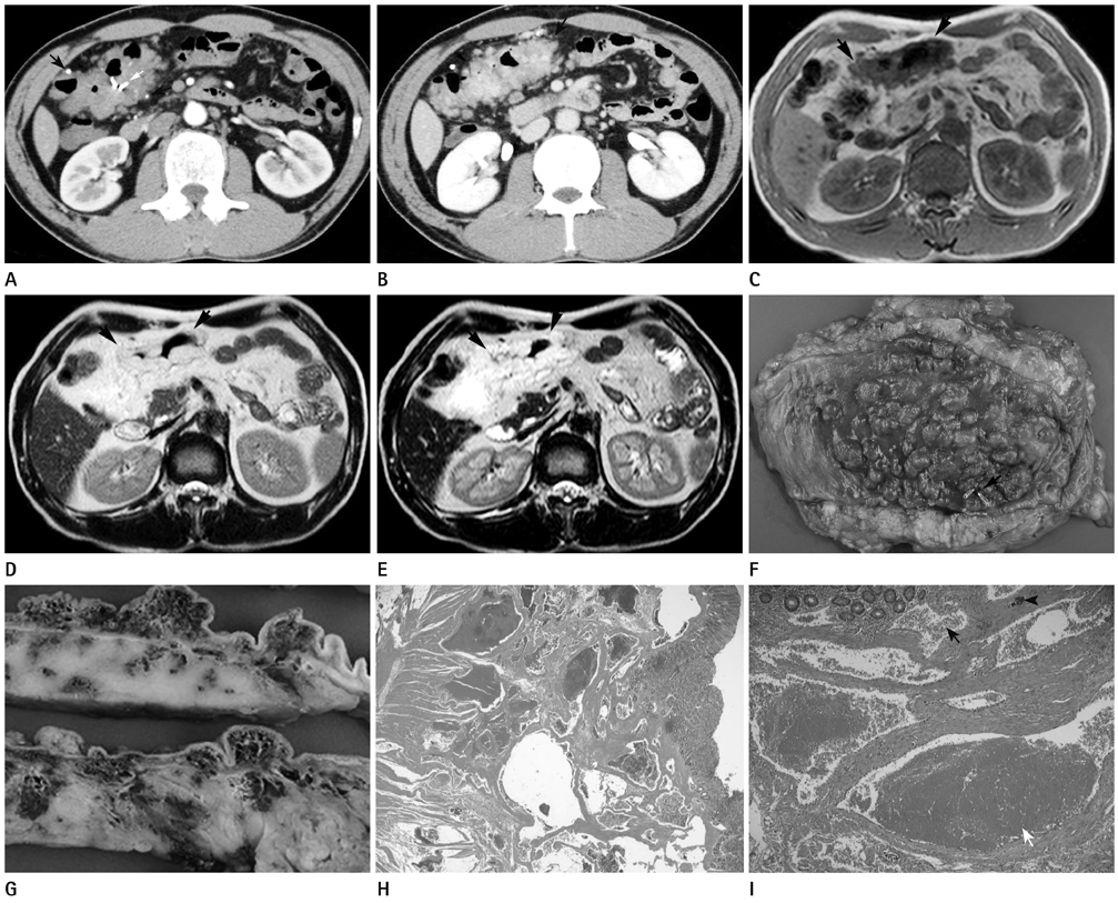
Fig. 1 Diffuse cavernous hemangioma of the transverse colon in a 41-year-old man. A. Arterial phase CT shows two small intralesional nodular calcifications (black arrow) and hemostatic clip (white arrow). B. Delayed phase CT shows circumferential wall thickening with heterogeneous nodular high enhancement in the transverse colon (black arrows). The same characteristic nodular and sperpigenous enhancements are noted in the pericolic fat. C-E. MR imaging shows thickened wall of the transverse colon (black arrows) with low signal intensity on T1-weighted image and high signal intensity on T2 and heavily T2 weighted image relative to that of the mesenteric fat. F. Gross pathological examination of transverse colonic mucosa shows huge, ill-defined, markedly congested, bluish purple, discrete to mulberry-like, conglomerated, submucosal tumefaction. A hemostatic clip is seen (black arrow). G. The cut sections of gross pathological examination show numerous, blood-filled, sponge-like, microcystic spaces, scattered at the submucosa, the muscularis propria and the pericolic adipose tissue. H. Histological examination of low power view shows multiple closely apposed multilocular blood-filled thin walled vascular channels, sharing thin fibrocollagenous common wall, at the submucosa and the muscularis propria (H&E, × 10). I. Histological examination of high power view shows multilocular cavernous vascular channels lined by bland-looking, single endothelial cells, often containing inraluminal, fresh (black arrow) or organizing (white arrow), fibrin thrombi and minimal focus of dystrophic calcification (black arrowhead) (H&E, × 100).
Reference
-
1. Londono-Schimmer EE, Ritchie JK, Hawley PR. Coloanal sleeve anastomosis in the treatment of diffuse cavernous haemangioma of the rectum: long-term results. Br J Surg. 1994; 81:1235–1237.2. Demircan O, Sönmez H, Zeren S, Cosşar E, Bicakci K, Ozkan S. Diffuse cavernous hemangioma of the rectum and sigmoid colon. Dig Surg. 1998; 15:713–715.3. Lyon DT, Mantia AG. Large-bowel hemangiomas. Dis Colon Rectum. 1984; 27:404–414.4. Tanaka N, Onda M, Seya T, Furukawa K, Kumazaki T. Diffuse cavernous haemangioma of the rectum. Eur J Surg. 1999; 165:280–283.5. Iafrate F, Laghi A, Paolantonio P, Rengo M, Mercantini P, Ferri M, et al. Preoperative staging of rectal cancer with MR Imaging: correlation with surgical and histopathologic findings. Radiographics. 2006; 26:701–714.6. Amarapurkar D, Jadliwala M, Punamiya S, Jhawer P, Chitale A, Amarapurkar A. Cavernous hemangiomas of the rectum: report of three cases. Am J Gastroenterol. 1998; 93:1357–1359.7. Tung GA, Vaccaro JP, Cronan JJ, Rogg JM. Cavernous hemangioma of the liver: pathologic correlation with high-field MR imaging. AJR Am J Roentgenol. 1994; 162:1113–1117.8. Ashida C, Fishman EK, Zerhouni EA, Herlong FH, Siegelman SS. Computed tomography of hepatic cavernous hemangioma. J Comput Assist Tomogr. 1987; 11:455–460.
