MRI Features of Atypical Cavernous Hemangioma Showing Central Filling Defect: A Case Report
- Affiliations
-
- 1Department of Radiology, Eulji University Hospital, Daejeon, Korea. midosyu@eulji.ac.kr
- KMID: 2407931
- DOI: http://doi.org/10.3348/jksr.2018.78.4.259
Abstract
- Cavernous hemangioma is a benign tumor composed of vascular structures and connective tissue. Typical imaging findings of cavernous sinus cavernous hemangioma are a well-defined contour-bulging mass, with homogeneous high signal intensity on T2-weighted images (T2WI), marked homogeneous enhancement of the cavernous sinus, and some sellar extension on magnetic resonance images. However, we experienced an unusual case of cavernous hemangioma, with central filling defects on delayed contrast-enhanced T1-weighted images and central, dark signal intensities on T2WI, which made the diagnosis difficult. The central portion of the lesion was pathologically consistent with central thrombosis. We present the clinical and imaging findings of this unusual case of cavernous hemangioma.
MeSH Terms
Figure
-
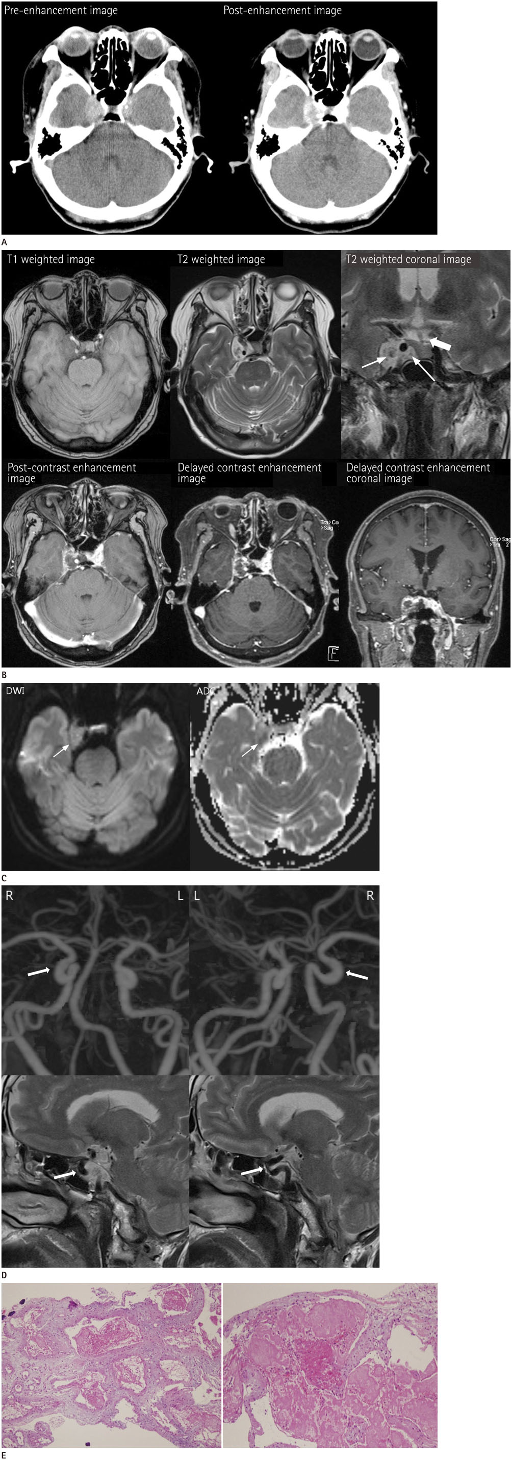
Fig. 1 A 67-year-old woman with a cavernous sinus mass with central filling defect on delayed contrast enhanced T1-weighted images. A. On axial pre-contrast CT image, the well-defined contour bulging mass in the right cavernous sinus shows high attenuation. Multiple-scattered, tiny, high attenuation foci located in the mass, suggest calcifications. On axial post-contrast CT image, the mass shows intense peripheral enhancement. Compared to the peripheral portion, the irregular central portion shows poor enhancement. CT = computed tomography B. The mass shows intermediate-to-low signal intensity on axial T1WI and high signal intensity with dark internal foci on axial T2WI. On coronal T2WI, dark portions are located at the internal and external aspects of the mass (thin arrows). Furthermore, the pituitary stalk is deviated to the left by the mass effect (thick arrow). On early post-contrast enhanced axial T1WI, the mass shows intense peripheral enhancement with a poorly enhancing inner portion. On delayed post-contrast-enhanced axial and coronal T1WI, taken as three-dimensional T1 gradient echo, the central portion shows no definite filling-in or delayed enhancement pattern. T1WI = T1-weighted image, T2WI = T2-weighted image C. The posterior portion of the mass, located in the right cavernous sinus, shows slightly high signal intensity on DWI and low signal intensity on ADC maps, meaning diffusion restriction (arrow). D. On computed tomography angiography (superior) and a sagittal T2-weighted images (inferior), the portion of the right internal carotid artery in the mass (arrows) shows no luminal narrowing or obstruction. ADC = apparent diffusion coefficient, DWI = diffusion weighted image E. Hematoxylin and eosin stain, × 100. Histologic specimen shows an internal thrombosis in the cavernous hemangioma.
Reference
-
1. Salanitri GC, Stuckey SL, Murphy M. Extracerebral cavernous hemangioma of the cavernous sinus: diagnosis with MR imaging and labeled red cell blood pool scintigraphy. AJNR Am J Neuroradiol. 2004; 25:280–284.2. Sohn CH, Kim SP, Kim IM, Lee JH, Lee HK. Characteristic MR imaging findings of cavernous hemangiomas in the cavernous sinus. AJNR Am J Neuroradiol. 2003; 24:1148–1151.3. Krief O, Sichez JP, Chedid G, Bencherif B, Zouaoui A, Le Bras F, et al. Extraaxial cavernous hemangioma with hemorrhage. AJNR Am J Neuroradiol. 1991; 12:988–990.4. Jinhu Y, Jianping D, Xin L, Yuanli Z. Dynamic enhancement features of cavernous sinus cavernous hemangiomas on conventional contrast-enhanced MR imaging. AJNR Am J Neuroradiol. 2008; 29:577–581.
Article5. Ahmadi J, Miller CA, Segall HD, Park SH, Zee CS, Becker RL. CT patterns in histopathologically complex cavernous hemangiomas. AJNR Am J Neuroradiol. 1985; 6:389–393.6. Savoiardo M, Strada L, Passerini A. Intracranial cavernous hemangiomas: neuroradiologic review of 36 operated cases. AJNR Am J Neuroradiol. 1983; 4:945–950.
- Full Text Links
-
- Actions
-
Cited
- CITED
-
- Close
- Share
-
- Similar articles
-
- Extracerebral Cavernous Hemangioma of the Middle Cranial Fossa: Report of 2 Cases and Review of Literature
- A Case of Cavernous Hemangioma of the Bladder
- A Case of Extradural Cavernous Hemangioma with Reuptured Disc
- Parenchymal Cavernous Hemangioma of the Breast showing Atypical Imaging Features: A Case Report
- Subcutaneous Cavernous Hemangioma of the Breast: A Case Report
