Tuberc Respir Dis.
2006 Jan;60(1):97-101.
Disseminated Coccidioidomycosis Presenting with Miliary Nodules
- Affiliations
-
- 1Department of Internal Medicine, College of Medicine, Korea University, Seoul, Korea. chol-shin@hanmail.net
Abstract
- Coccidioidomycosis is a rare systemic fungal infection in Korea. However, the incidence of coccidioidomycosis has recently begun to increase due to the increasing incidence of people traveling overseas to endemic areas. In previously reported cases of coccidioidomycosis in Korea, the radiographic findings usually showed a solitary pulmonary nodule, pleural effusion, cavitation, and hilar lymphadenopathy, but no miliary nodules. We report a case of disseminated coccidioidomycosis with miliary nodules in an immunocompetent patient. A 32 year old male, who had traveled in Corona, New Mexico, USA, was admitted for an evaluation of persistent cough with fever. Chest radiography revealed initially diffuse multiple small nodules that appeared to be miliary tuberculosis. However, a subsequent evaluation revealed that he had disseminated coccidioidomycosis.
Keyword
MeSH Terms
Figure
-
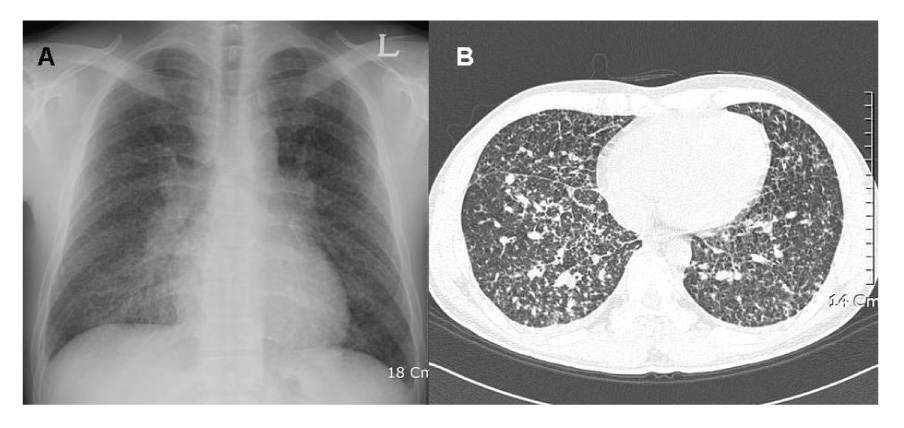
Figure 1 Chest radiography (A) and high resolution computed tomography (B) on admission showed diffuse, multiple, and small nodules with interlobular septal thickening on both whole lung field.
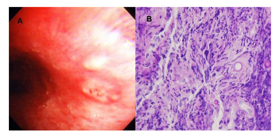
Figure 2 Bronchoscopic examination showed whitish, nodular lesion on bronchial mucosa of left main bronchus (A) and histologic findings of specimens obtained by bronchoscopic biopy (B) showed a marked eosinophilic infiltration and a few spherules (hematoxylin and eosin, ×200).

Figure 3 Pustules on facial skin (A) and histologic findings of specimens obtained by excisional skin biopy (B) showed chronic granulomatous inflammation with thick-walled mature spherules containing endospores (hematoxylin and eosin, ×400).
Reference
-
1. Kim JH, Kim MA, Yang SK, Choi TY, Kim CW, Kim KH. A case of coccidioidomycosis. Korean J Dermatol. 1976. 14:73–79.2. Yang HS, Lee J, Lim CM, Lee SD, Koh Y, Kim WS, et al. A case of coccidioidomycosis manifested as solitary pulmonary nodule. Tuberc Respir Dis. 1999. 46:266–272.3. Shin JS, Lee IS, Shin C, Kim A. Pulmonary coccidioidomycosis in an immigrant. Tuberc Respir Dis. 2001. 51:448–452.4. Lim G, Woo J, Chung YT, Uh ST, Park CS. A case of pulmonary coccidioidomycosis. Tuberc Respir Dis. 1990. 37:440–444.5. Galgiani JN. Coccidioidomycosis: a regional disease of national importance. Ann Intern Med. 1999. 130:293–300.6. Stevens DA. Coccidioidomycosis. N Engl J Med. 1995. 332:1077–1082.7. Chuang A, Thomas R, Hoffman RS. Disseminated coccidioidomycosis in an immunocompetent person living in New York city. J Urban Health. 2005. 82:339–345.8. Feldman BS, Snyder LS. Primary pulmonary coccidioidomycosis. Semin Respir Infect. 2001. 16:231–237.9. Arsura EL, Kilgore WB. Miliary coccidioidomycosis in the immunocompetent. Chest. 2000. 117:404–409.10. Polesky A, Kirsch CM, Snyder LS, LoBue P, Kagawa FT, Dykstra BJ, et al. Airway coccidioidomycosis: report of cases and review. Clinic Infect Dis. 1999. 28:1273–1280.11. Arnold MG, Arnold JC, Bloom DC, Brewster DF, Thiringer JK. Head and neck manifestations of disseminated coccidioidomycosis. Laryngoscope. 2004. 114:747–752.12. Galgiani JN, Ampel NM, Catanzaro A, Johnson RH, Stevens DA, Williams PL. Practice guidelines for the treatment of coccidioidomycosis. Clin Infect Dis. 2000. 30:658–661.13. Deresinski SC. Coccidioidomycosis: efficacy of new agents and future prospects. Curr Opin Infect Dis. 2001. 14:693–696.
- Full Text Links
-
- Actions
-
Cited
- CITED
-
- Close
- Share
-
- Similar articles
-
- A Case of Disseminated Coccidioidomycosis Confirmed by Skin Biopsy in Korea
- A case of disseminated coccidioidomycosis: autopsy report
- Disseminated Coccidioidomycosis with Cutaneous Manifestation
- Diffuse Large B-Cell Lymphoma Manifesting as Miliary Nodules in the Lung: A Case Report
- A Case of Disseminated Coccidioidomycosis Involving the Lymph Nodes, the Skin, and the Brain
