Endodontic treatment of a C-shaped mandibular second premolar with four root canals and three apical foramina: a case report
- Affiliations
-
- 1Division of Endodontics, Columbia University College of Dental Medicine, New York, NY, USA. sgk2114@columbia.edu
- KMID: 2316966
- DOI: http://doi.org/10.5395/rde.2016.41.1.68
Abstract
- This case report describes a unique C-shaped mandibular second premolar with four canals and three apical foramina and its endodontic management with the aid of cone-beam computer tomography (CBCT). C-shaped root canal morphology with four canals was identified under a dental operating microscope. A CBCT scan was taken to evaluate the aberrant root canal anatomy and devise a better instrumentation strategy based on the anatomy. All canals were instrumented to have a 0.05 taper using 1.0 mm step-back filing with appropriate apical sizes determined from the CBCT scan images and filled using a warm vertical compaction technique. A C-shaped mandibular second premolar with multiple canals is an anatomically rare case for clinicians, yet its endodontic treatment may require a careful instrumentation strategy due to the difficulty in disinfecting the canals in the thin root area without compromising the root structure.
Keyword
MeSH Terms
Figure
-
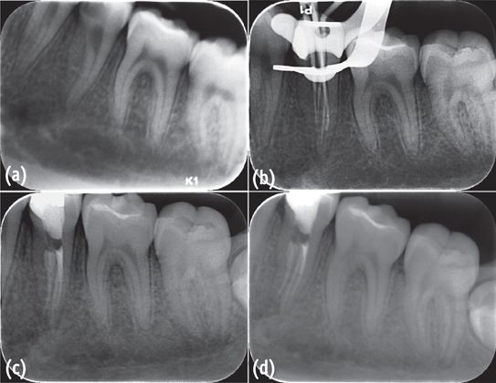
Figure 1 Root canal treatment of the C-shaped left mandibular second premolar with four canals and three apical foramina. (a) A preoperative periapical radiograph; (b) Master cone fit; (c, d) Postoperative periapical radiographs showing mesial and mesiobuccal canals joining in the coronal third of the root.
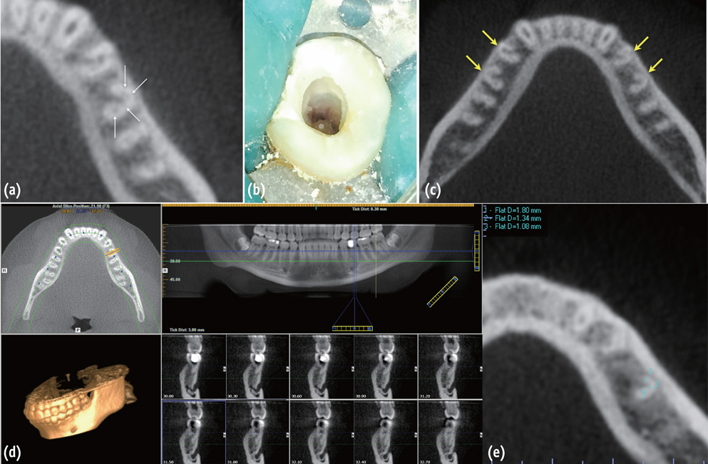
Figure 2 A CBCT scan and access cavity of the C-shaped left mandibular second premolar. (a) Axial view showing four canals in the C-shaped left mandibular second premolar; (b) Occlusal view of access cavity showing a C-shaped root canal configuration; (c) Axial view showing bilateral C-shaped mandibular first and second premolars; (d) Axial, panoramic, and sagittal views showing a fused root configuration of the left mandibular second premolar; (e) Axial view showing a measurement of root thicknesses around canals. The root thicknesses were measured individually at all levels with 0.3 mm interval from each canal to the outer surface of the root, and from the apical foramina to the canal orifices.
Reference
-
1. Pineda F, Kuttler Y. Mesiodistal and buccolingual roentgenographic investigation of 7,275 root canals. Oral Surg Oral Med Oral Pathol. 1972; 33:101–110.
Article2. Sert S, Bayirli GS. Evaluation of the root canal configurations of the mandibular and maxillary permanent teeth by gender in the Turkish population. J Endod. 2004; 30:391–398.
Article3. Awawdeh LA, Al-Qudah AA. Root form and canal morphology of mandibular premolars in a Jordanian population. Int Endod J. 2008; 41:240–248.
Article4. Zillich R, Dowson J. Root canal morphology of mandibular first and second premolars. Oral Surg Oral Med Oral Pathol. 1973; 36:738–744.
Article5. Holtzman L. Root canal treatment of mandibular second premolar with four root canals: a case report. Int Endod J. 1998; 31:364–366.
Article6. Bram SM, Fleisher R. Endodontic therapy in a mandibular second bicuspid with four canals. J Endod. 1991; 17:513–515.7. Rhodes JS. A case of unusual anatomy: a mandibular second premolar with four canals. Int Endod J. 2001; 34:645–648.
Article8. Macri E, Zmener O. Five canals in a mandibular second premolar. J Endod. 2000; 26:304–305.
Article9. Sachdeva GS, Ballal S, Gopikrishna V, Kandaswamy D. Endodontic management of a mandibular second premolar with four roots and four root canals with the aid of spiral computed tomography: a case report. J Endod. 2008; 34:104–107.
Article10. Yu X, Guo B, Li KZ, Zhang R, Tian YY, Wang H, Hu T. Cone-beam computed tomography study of root and canal morphology of mandibular premolars in a western Chinese population. BMC Med Imaging. 2012; 12:18.
Article11. Rahimi S, Shahi S, Yavari HR, Manafi H, Eskandarzadeh N. Root canal configuration of mandibular first and second premolars in an Iranian population. J Dent Res Dent Clin Dent Prospects. 2007; 1:59–64.12. Jafarzadeh H, Wu YN. The C-shaped root canal configuration: a review. J Endod. 2007; 33:517–523.
Article13. Lu TY, Yang SF, Pai SF. Complicated root canal morphology of mandibular first premolar in a Chinese population using the cross section method. J Endod. 2006; 32:932–936.
Article14. Sidow SJ, West LA, Liewehr FR, Loushine RJ. Root canal morphology of human maxillary and mandibular third molars. J Endod. 2000; 26:675–678.
Article15. Cleghorn BM, Christie WH, Dong CC. Root and root canal morphology of the human permanent maxillary first molar: a literature review. J Endod. 2006; 32:813–821.
Article16. Manning SA. Root canal anatomy of mandibular second molars. Part II. C-shaped canals. Int Endod J. 1990; 23:40–45.17. Barnett F. Mandibular molar with C-shaped canal. Endod Dent Traumatol. 1986; 2:79–81.
Article18. Varrela J. Effect of 45,X/46,XX mosaicism on root morphology of mandibular premolars. J Dent Res. 1992; 71:1604–1606.
Article19. Varrela J. Root morphology of mandibular premolars in human 45,X females. Arch Oral Biol. 1990; 35:109–112.
Article20. Kusiak A, Sadlak-Nowicka J, Limon J, Kochńska B. Root morphology of mandibular premolars in 40 patients with Turner syndrome. Int Endod J. 2005; 38:822–826.
Article21. Chauhan R, Singh S, Chandra A. A rare occurrence of bilateral C-shaped roots in mandibular first and second premolars diagnosed with the aid of spiral computed tomography. J Clin Exp Dent. 2014; 6:e440–e443.
Article22. Shah DY. C-shaped root canal configuration in mandibular second premolar: report of an unusual case and its endodontic management. J Int Clin Dent Res Organ. 2012; 4:18–20.
Article23. Hunter AJ, Feiglin B, Williams JF. Effects of post placement on endodontically treated teeth. J Prosthet Dent. 1989; 62:166–172.
Article24. Tilk MA, Lommel TJ, Gerstein H. A study of mandibular and maxillary root widths to determine dowel size. J Endod. 1979; 5:79–82.
Article25. Johnson JK, Schwartz NL, Blackwell RT. Evaluation and restoration of endodontically treated posterior teeth. J Am Dent Assoc. 1976; 93:597–605.
Article26. Lim SS, Stock CJ. The risk of perforation in the curved canal: anticurvature filing compared with the stepback technique. Int Endod J. 1987; 20:33–39.
Article27. Zuckerman O, Katz A, Pilo R, Tamse A, Fuss Z. Residual dentin thickness in mesial roots of mandibular molars prepared with Lightspeed rotary instruments and Gates-Glidden reamers. Oral Surg Oral Med Oral Pathol Oral Radiol Endod. 2003; 96:351–355.
Article28. Jerome CE. C-shaped root canal systems: diagnosis, treatment, and restoration. Gen Dent. 1994; 42:424–427.29. Cooke HG 3rd, Cox FL. C-shaped canal configurations in mandibular molars. J Am Dent Assoc. 1979; 99:836–839.
Article30. Haddad GY, Nehme WB, Ounsi HF. Diagnosis, classification, and frequency of C-shaped canals in mandibular second molars in the Lebanese population. J Endod. 1999; 25:268–271.
Article
- Full Text Links
-
- Actions
-
Cited
- CITED
-
- Close
- Share
-
- Similar articles
-
- Root canal treatment of a mandibular second premolar with three separate root canals
- Use of cone-beam computed tomography and three-dimensional modeling for assessment of anomalous pulp canal configuration: a case report
- Outcome Assessment of Endodontic Treatment of Mandibular Second Molars with C-shaped Canals in Elderly Patients
- A retrospective study on incidence of C-shaped canals in mandibular second molars
- Healing outcomes of root canal treatment for C-shaped mandibular second molars: a retrospective analysis
