Healing after horizontal root fractures: 3 cases with 2-year follow-up
- Affiliations
-
- 1Department of Conservative Dentistry, Wonkwang University School of Dentistry, Iksan, Korea. conspsj@wonkwang.ac.kr
- 2Department of Conservative Dentistry, Chonbuk National University School of Dentistry, Jeonju, Korea.
Abstract
- Among dental traumas, horizontal root fractures are relatively uncommon injuries. Proper initial management and periodical evaluation is essential for the successful treatment of a root-fractured tooth. If pulpal necrosis develops, endodontic treatment is indicated, exclusively for the coronal fragment. Fragment diastases exert a great influence on healing at the fracture line and on pulpal necrosis. An adequately treated root-fractured tooth has a good prognosis. This case report describes the treatment and 2-yr follow up of 3 maxillary central incisors, first with horizontal root fracture, second with horizontal root fracture and avulsion, and third with horizontal root fracture and lateral luxation. All three cases were treated with mineral trioxide aggregate (ProRoot, Dentsply). During 2 yr of follow-up evaluation, the root-fractured teeth of the present patients were well retained in the arch, showing periodontal healing, even after endodontic treatment.
Figure
-
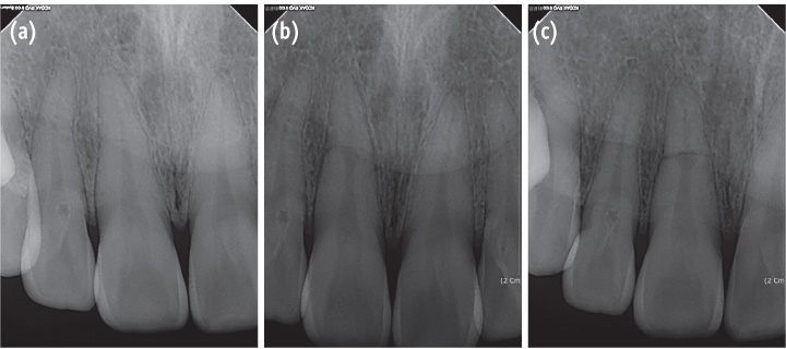
Figure 1 (a) The radiograph of the initial visit shows a horizontal root fracture line on the maxillary right central incisor; (b) The 4-week follow-up radiograph shows internal resorption around the fracture line; (c) The 6-week follow-up radiograph shows that the internal resorption site is larger and more distinct.
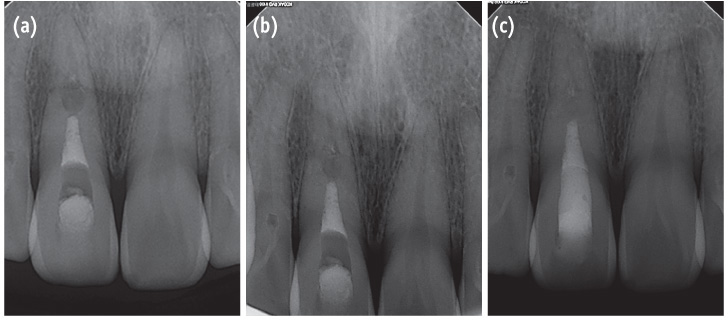
Figure 2 Radiograph of the tooth. (a) After root canal treatment; (b) At the 1-year follow-up evaluation; (c) At the 2-year follow-up evaluation. 2-year follow-up radiograph reveals healing with the interposition of hard tissue between the fragments.

Figure 3 Radiograph of the right maxillary central incisor with horizontal root fracture. (a) After repositioning and applying a resin-wire splint; (b) After root canal treatment; (c) At the 2-year follow-up evaluation. 2-year follow-up radiograph reveals healing with the interposition of hard and soft tissue between the fragments.
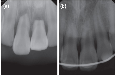
Figure 4 Radiographs of the emergency visit shows the teeth. (a) Before the application of the resin-wire splint; (b) After repositioning and the application of the resin-wire splint.

Figure 5 Images were obtained 3 weeks after the trauma. (a) The periapical radiograph shows a radiolucent lesion around the fracture line on tooth #21; (b) The clinical photograph shows narrow and deep pocket depth and gingival swelling on tooth #21.

Figure 6 Radiograph of the teeth. (a) After the root canal treatment; (b) At the 1-year follow-up evaluation; (c) at the 2-year follow-up evaluation. The radiolucent lesion has disappeared on tooth #21. 2-year follow-up radiograph reveals healing with interposition of soft tissue between the fragments.
Reference
-
1. Andreasen FM, Andreasen JO. Root fractures. In : Andreasen JO, Andreasen FM, editors. Textbook and color atlas of traumatic injuries to the teeth. 3rd ed. Copenhagen: Munksgaard;1994. p. 279–314.2. Clark SJ, Eleazer P. Management of a horizontal root fracture after previous root canal therapy. Oral Surg Oral Med Oral Pathol Oral Radiol Endod. 2000; 89:220–223.
Article3. Cvek M, Mejàre I, Andreasen JO. Conservative endodontic treatment of teeth fractured in the middle or apical part of the root. Dent Traumatol. 2004; 20:261–269.
Article4. Cvek M, Tsilingaridis G, Andreasen JO. Survival of 534 incisors after intra-alveolar root fracture in patients aged 7-17 years. Dent Traumatol. 2008; 24:379–387.
Article5. Andrade ES, de Campos Sobrinho AL, Andrade MG, Matos JL. Root healing after horizontal fracture: a case report with a 13-year follow up. Dent Traumatol. 2008; 24:e1–e3.
Article6. Versiani MA, de Sousa CJ, Cruz-Filho AM, Perez DE, Sousa-Neto MD. Clinical management and subsequent healing of teeth with horizontal root fractures. Dent Traumatol. 2008; 24:136–139.
Article7. Andreasen FM, Andreasen JO. Resorption and mineralization processes following root fracture of permanent incisors. Endod Dent Traumatol. 1988; 4:202–214.
Article8. Andreasen JO, Andreasen FM, Mejàre I, Cvek M. Healing of 400 intra-alveolar root fractures. 2. Effect of treatment factors such as treatment delay, repositioning, splinting type and period and antibiotics. Dent Traumatol. 2004; 20:203–211.
Article9. Hargreaves KM, Cohen S. Cohen's pathways of the pulp. 10th ed. St. Louis: Mosby Elsevier;2010. p. 635–637.10. Shin JH, Kim RJ. Management of horizontal root fractures by fabrication of canine protected occlusion using composite resin. Restor Dent Endod. 2012; 37:180–184.
Article11. Wölner-Hanssen AB, von Arx T. Permanent teeth with horizontal root fractures after dental trauma. A retrospective study. Schweiz Monatsschr Zahnmed. 2010; 120:200–212.12. Andreasen JO, Hjorting-Hansen E. Intra-alveolar root fractures: radiographic and histologic study of 50 cases. J Oral Surg. 1967; 25:414–426.13. Andreasen JO, Andreasen FM, Mejàre I, Cvek M. Healing of 400 intra-alveolar root fractures. 1. Effect of preinjury and injury factors such as sex, age, stage of root development, fracture type, location of fracture and severity of dislocation. Dent Traumatol. 2004; 20:192–202.
Article
- Full Text Links
-
- Actions
-
Cited
- CITED
-
- Close
- Share
-
- Similar articles
-
- Management of horizontal root fractures by fabrication of canine protected occlusion using composite resin
- Use of mineral trioxide aggregate in the treatment of horizontal root fracture with a 4-year follow-up: case report
- Factors Affecting the Pulp and Root Healing of Root Fractures in Immature Permanent Teeth
- Treatment of horizontal root-fractured maxillary incisors
- Pulp revascularization with and without platelet-rich plasma in two anterior teeth with horizontal radicular fractures: a case report
