Korean J Urol.
2012 Mar;53(3):159-164.
Characterization of Small Renal Masses Less than 4 cm with Quadriphasic Multidetector Helical Computed Tomography: Differentiation of Benign and Malignant Lesions
- Affiliations
-
- 1Department of Urology, Kyung Hee University School of Medicine, Seoul, Korea. juro@khu.ac.kr
Abstract
- PURPOSE
To identify the characteristic quadriphasic (unenhanced, corticomedullary, nephrographic, and excretory phase) helical multidetector computed tomography (MDCT) features of renal masses less than 4 cm to distinguish benign from malignant renal masses.
MATERIALS AND METHODS
In total, 84 patients were retrospectively analyzed to determine the characteristic features for the prediction of subtypes of small renal masses. The patients' age, gender, and tumor size and CT features, including the presence of intra-tumor degenerative changes, septation, calcification, and wall irregularity, were evaluated. In addition, the degree and pattern of enhancement obtained during four phases were analyzed. The relationship between the subtype of the small renal masses and the gender, morphological features, and pattern of contrast enhancement on the CT was analyzed by using the chi-square test. Tumor size and degree of contrast enhancement were compared by the Mann-Whitney U test. The predictive value of each of the CT features was determined by multivariate logistic regression analysis.
RESULTS
Of the 84 small renal masses, 17 (20%) were benign and 67 (80%) were malignant. Univariate analysis revealed that renal cell carcinoma lesions showed heterogeneous enhancement (p=0.002) and higher mean attenuation value on the corticomedullary and nephrographic phases (135.1+/-53.9, p=0.000, and 132.4+/-43.6, p=0.006). The multivariate analysis with logistic regression model showed that only the mean attenuation value on the corticomedullary phase had a statistically significant correlation (p=0.021).
CONCLUSIONS
For the characterization of small renal masses, the degree of enhancement on the corticomedullary phase is a valuable parameter. Furthermore, the heterogeneous enhancement pattern and degree of enhancement on the nephrographic phase can provide information for differentiating small renal masses.
MeSH Terms
Figure
-
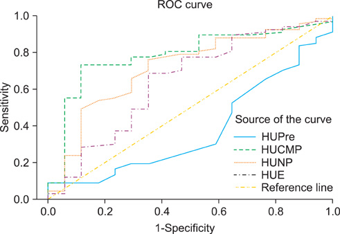
FIG. 1 Receiver operating characteristic (ROC) curves for the degree of contrast enhancement as a distinguishing factor between small malignant masses and small benign masses. The area under the curve for the corticomedullary phase (CMP) was 0.785 (95% confidence interval [CI], 0.667 to 0.902), which was statistically significant, and that for the nephrographic phase (NP) was 0.717 (95% CI, 0.579 to 0.854). CMP, corticomedullary phase; NP, nephrographic phase.
Reference
-
1. Catalano C, Fraioli F, Laghi A, Napoli A, Pediconi F, Danti M, et al. High-resolution multidetector CT in the preoperative evaluation of patients with renal cell carcinoma. AJR Am J Roentgenol. 2003. 180:1271–1277.2. Gakis G, Kramer U, Schilling D, Kruck S, Stenzl A, Schlemmer HP. Small renal oncocytomas: differentiation with multiphase CT. Eur J Radiol. 2011. 80:274–278.3. Bird VG, Kanagarajah P, Morillo G, Caruso DJ, Ayyathurai R, Leveillee R, et al. Differentiation of oncocytoma and renal cell carcinoma in small renal masses (<4 cm): the role of 4-phase computerized tomography. World J Urol. 2011. 29:787–792.4. Sheth S, Scatarige JC, Horton KM, Corl FM, Fishman EK. Current concepts in the diagnosis and management of renal cell carcinoma: role of multidetector CT and three-dimensional CT. Radiographics. 2001. 21 Spec No:S237–S254.5. Kopka L, Fischer U, Zoeller G, Schnidt C, Ringert RH, Grabbe E. Dual-phase helical Ct of the kidney: value of the corticomedullary and nephrographic phase for evaluation of renal lesions and preoperative staging of renal cell carcinoma. AJR Am J Roentgenol. 1997. 169:1573–1578.6. Silver DA, Morash C, Brenner P, Campbell S, Russo P. Pathologic findings at the time of nephrectomy for renal mass. Ann Surg Oncol. 1997. 4:570–574.7. Ozen H, Colowick A, Freiha FS. Incidentally discovered solid renal masses: what are they? Br J Urol. 1993. 72:274–276.8. Kutikov A, Fossett LK, Ramchandani P, Tomaszewski JE, Siegelman ES, Banner MP, et al. Incidence of benign pathologic findings at partial nephrectomy for solitary renal mass presumed to be renal cell carcinoma on preoperative imaging. Urology. 2006. 68:737–740.9. Schachter LR, Cookson MS, Chang SS, Smith JA Jr, Dietrich MS, Jayaram G, et al. Second prize: frequency of benign renal cortical tumors and histologic subtypes based on size in a contemporary series: what to tell our patients. J Endourol. 2007. 21:819–823.10. Duchene DA, Lotan Y, Cadeddu JA, Sagalowsky AI, Koeneman KS. Histopathology of surgically managed renal tumors: analysis of a contemporary series. Urology. 2003. 62:827–830.11. Landis SH, Murray T, Bolden S, Wingo PA. Cancer statistics, 1999. CA Cancer J Clin. 1999. 49:8–31.12. Kim JK, Kim TK, Ahn HJ, Kim CS, Kim KR, Cho KS. Differentiation of subtypes of renal cell carcinoma on helical CT scan. AJR Am J Roentgenol. 2002. 178:1499–1506.13. Jinzaki M, Tanimoto A, Mukai M, Ikeda E, Kobayashi S, Yuasa Y, et al. Double-phase helical CT of small renal parenchymal neoplasms: correlation with pathologic findings and tumor angiogenesis. J Comput Assist Tomogr. 2000. 24:835–842.14. Choudhary S, Rajesh A, Mayer NJ, Mulcahy KA, Haroon A. Renal oncocytoma: CT features cannot reliably distinguish oncocytoma from other renal neoplasms. Clin Radiol. 2009. 64:517–522.15. Davidson AJ, Hayes WS, Hartman DS, McCarthy WF, Davis CJ Jr. Renal oncocytoma and carcinoma: failure of differentiation with CT. Radiology. 1993. 186:693–696.16. Hélénon O, Chrétien Y, Paraf F, Melki P, Denys A, Moreau JF. Renal cell carcinoma containing fat: demontration with CT. Radiology. 1993. 188:429–430.17. Strotzer M, Lehner KB, Becker K. Detection of fat in a renal cell carcinoma mimicking angiomyolipoma. Radiology. 1993. 188:427–428.18. Kim JK, Park SY, Shon JH, Cho KS. Angiomyolipoma with minimal fat: differentiation from renal cell carcinoma at biphasic helical CT. Radiology. 2004. 230:677–684.19. Kim JY, Kim JK, Kim N, Cho KS. CT histogram analysis: differentiation of angiomyolipoma without visible fat from renal cell carcinoma at CT imaging. Radiology. 2008. 246:472–479.20. Garant M, Bonaldi VM, Taourel P, Pinsky MF, Bret PM. Enhancement patterns of renal masses during multiphase helical CT acquistions. Abdom imaging. 1998. 23:431–436.21. Israel GM, Bosniak MA. Renal imaging for diagnosis and staging of renal cell carcinoma. Urol Clin North Am. 2003. 30:499–514.22. Szolar DH, Kammerhuber F, Altziebler S, Tillich M, Breinl E, Fotter R, et al. Multiphasic helical CT of the kidney: increased conspicuity for detection and characterization of small (<3-cm) renal masses. Radiology. 1997. 202:211–217.
- Full Text Links
-
- Actions
-
Cited
- CITED
-
- Close
- Share
-
- Similar articles
-
- The Clinical Significance of Bosniak Classification in Cystic Renal Masses : Usefulness of Preoperative Computerized Tomography in Cystic Renal Masses
- Incidental Solid Renal Masses: Radiologic Assessment and Managements
- Differential Diagnosis of Adrenal Mass Using Imaging Modality: Special Emphasis on F-18 Fluoro-2-Deoxy-D-Glucose Positron Emission Tomography/Computed Tomography
- Usefulness of the Bosniak Classification in Cystic Renal Mass on CT
- The Comparison between Pre- and Postoperative Diagnosis in Renal Masses Smaller than 3cm in Diameter
