Diagnosing Acute Pyelonephritis with CT, (99m)Tc-DMSA SPECT, and Doppler Ultrasound: A Comparative Study
- Affiliations
-
- 1Department of Urology, College of Medicine, The Catholic University of Korea, Seoul, Korea. ysleemd@catholic.ac.kr
- 2Department of Radiology, College of Medicine, The Catholic University of Korea, Seoul, Korea.
Abstract
- PURPOSE
With growing interest in early imaging, the aim of our study was to define the most practical modality for routine clinical use for the diagnosis of acute pyelonephritis (APN). We compared the sensitivity of enhanced computerized tomography (CT), dimercaptosuccinic acid (DMSA) scintigraphy, and Doppler ultrasonography (DUS) by using clinical findings as the standard of reference. MATERIALS AND METHODS: A total of 207 APN patients (191 women, 16 men; mean age, 49.4 years; range, 17-88 years) were enrolled in this study. All the patients underwent imaging modalities during hospitalization. SPECT images were obtained 4 hours after injection of (99m)Tc-DMSA. Transverse and coronary CT images were obtained before and after injection of the contrast agent. DUS was performed in the longitudinal, transverse, and coronal planes. All the images were read independently by a single radiologist and a nuclear medicine specialist. The sensitivity of each modality for detecting APN was compared. RESULTS: CT showed significantly superior sensitivity compared with that of DUS (81.0% vs. 33.3%, respectively, n=147). DMSA scintigraphy also showed significantly superior sensitivity compared with that of DUS (74.7% vs. 33.3%, respectively, n=150). Compared with DMSA scintigraphy, CT showed superior sensitivity, but the difference was not statistically significant (81.0% vs. 74.8%, respectively, n=147, p=0.163). CONCLUSIONS: For cases of clinically suspected APN, CT and DMSA scintigraphy appear to be equally sensitive and reliable for detecting APN, although CT is more practical in various fields. DUS was significantly less sensitive.
Keyword
MeSH Terms
Figure
-
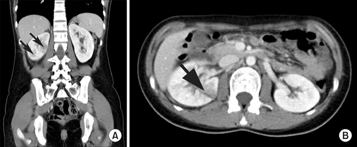
FIG. 1 (A) The coronary spiral CT scan obtained after intravenous administration of contrast agent demonstrates well-defined foci (arrows) of decreased attenuation in the lower pole of the right kidney. (B) A large area of parenchymal hypo-attenuation in the posterior aspect of the right kidney (arrow).

FIG. 2 (A) Dimercaptosuccinic acid (DMSA) scintigraphy demonstrates decreased uptake in the upper half and the lower portion of the right kidney (arrows). (B) The transverse images show a more accurate area of decreased uptake in the right kidney, especially in the anterior and the posterior aspect of the right kidney (arrows).
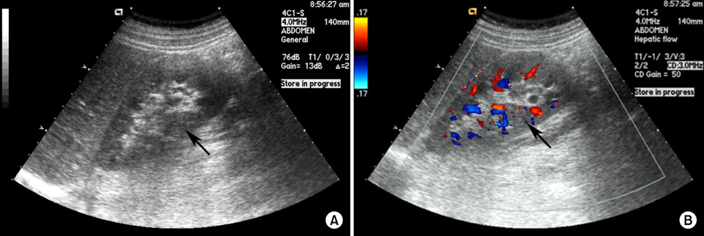
FIG. 3 (A) Renal Doppler ultrasonography (DUS) shows mild swelling and a wedge-shaped hypoechoic focus (arrow) of the right kidney related to acute pyelonephritis. (B) The color flow DUS image demonstrates diminished flow through the involved area (arrow).
Reference
-
1. Stunell H, Buckley O, Feeney J, Geoghegan T, Browne RF, Torreggiani WC. Imaging of acute pyelonephritis in the adult. Eur Radiol. 2007. 17:1820–1828.2. Kawashima A, Sandler CM, Goldman SM. Imaging in acute renal infection. BJU Int. 2000. 86:Suppl 1. 70–79.3. Majd M, Rushton HG, Jantausch B, Wiedermann BL. Relationship among vesicoureteral reflux P-fimbriated Escherichia coli and acute pyelonephritis in children with febrile urinary tract infection. Radiology. 1991. 119:578–585.4. Kawashima A, LeRoy AJ. Radiologic evaluation of patients with renal infections. Infect Dis Clin North Am. 2003. 17:433–456.5. Johansen TE. The role of imaging in urinary tract infections. World J Urol. 2004. 22:392–398.6. Shen Y, Brown MA. Renal Imaging in pyelonephritis. Nephrology. 2004. 9:22–25.7. Soulen MC, Fishman EK, Goldman SM, Gatewood OM. Bacterial renal infection: role of CT. Radiology. 1989. 171:703–707.8. Dacher JN, Boillot B, Eurin D, Marguet C, Mitrofanoff P, Le Dosseur P. Rational use of CT in acute pyelonephritis: findings and relationships with reflux. Pediatr Radiol. 1993. 23:281–285.9. Kass EJ, Fink-Bennett D, Cacciarelli AA, Balon H, Pavlock S. The sensitivity of renal scintigraphy and sonography in detecting nonobstructive acute pyelonephritis. J Urol. 1992. 148:606–608.10. Rushton HG, Majd M. Dimercaptosuccinic acid renal scintigraphy for the evaluation of pyelonephritis and scarring: a review of experimental and clinical studies. J Urol. 1992. 148:1726–1732.11. Kovanlikaya A, Okkay N, Cakmakci H, Ozdoðan O, Degirmenci B, Kavukcu S. Comparison of MRI and renal cortical scintigraphy findings in childhood acute pyelonephritis: preliminary experience. Eur J Radiol. 2004. 49:76–80.12. Dacher JN, Pfister C, Monroc M, Eurin D, LeDosseur P. Power Doppler sonographic pattern of acute pyelonephritis in children: comparison with CT. AJR Am J Roentgenol. 1996. 166:1451–1455.13. Hoddick W, Jeffrey RB, Goldberg HI, Federle MP, Laing FC. CT and sonography of severe renal and perirenal infections. AJR Am J Roentgenol. 1983. 140:517–520.14. June CH, Browning MD, Smith LP, Wenzel DJ, Pyatt RS, Checchio LM, et al. Ultrasonography and computed tomography in severe urinary tract infection. Arch Intern Med. 1985. 145:841–845.15. Majd M, Nussbaum Blask AR, Markle BM, Shalaby-Rana E, Pohl HG, Park JS, et al. Acute pyelonephritis: comparison of diagnosis with 99mTc-DMSA, SPECT, spiral CT, MR imaging, and power Doppler US in an experimental pig model. Radiology. 2001. 218:101–108.16. Safrin S, Siegel D, Black D. Pyelonephritis in adult women: inpatient versus outpatient therapy. Am J Med. 1988. 85:793–798.17. Chung JM, Choi S, Lee SD. Affecting factors on the treatment of acute pyelonephritis. Korean J Urogenital Tract Infect Inflamm. 2007. 2:73–77.18. Jung YH, Cho IR, Lee SE, Lee KC, Kim JG, Jeon JS, et al. Comparative analysis of clinical parameters in acute pyelonephritis. Korean J Urol. 2007. 48:29–34.19. Bang SH, Chang IH, Han JH, Ahn SH. C-reactive protein is a useful marker to predict the severity and early response of acute pyelonephritis in women. Korean J Urol. 2007. 48:1143–1148.20. Han CH, Cho SY, Kang SH. Age-related radiological imaging in children with acute pyelonephritis. Korean J Urol. 2003. 44:780–784.21. Rauschkolb EN, Sandler CM, Patel S, Childs TL. Computed tomography of renal inflammatory disease. J Comput Assist Tomogr. 1982. 6:502–506.22. Rigsby CM, Rosenfield AT, Glickman MG, Hodson J. Hemorrhagic focal bacterial nephritis: findings on gray-scale sonography and CT. AJR Am J Roentgenol. 1986. 146:1173–1177.23. Soulen MC, Fishman EK, Goldman SM. Sequelae of acute renal infections: CT evaluation. Radiology. 1989. 173:423–426.24. Fraser IR, Birch D, Fairley KF, John S, Lichtenstein M, Tress B, et al. A prospective study of cortical scarring in acute febrile pyelonephritis in adults: clinical and bacteriological characteristics. Clin Nephrol. 1995. 43:159–164.25. Clautice-Engle T, Jeffrey RB Jr. Renal hypoperfusion: value of power Doppler imaging. AJR Am J Roentgenol. 1997. 168:1227–1231.26. Winters WD. Power Doppler sonographic evaluation of acute pyelonephritis in children. J Ultrasound Med. 1996. 15:91–96.
- Full Text Links
-
- Actions
-
Cited
- CITED
-
- Close
- Share
-
- Similar articles
-
- Power Doppler Sonography in Children with Acute Pyelonephritis
- Power Doppler Sonography for the Upper Urinary Tract Infection in Children
- Limitations of 99mTc-DMSA scan in diagnosing acute pyelonephritis in children
- Comparison of 99mTc-DMSA Renal Scan and Power Doppler Ultrasonography for the Detection of Acute Pyelonephritis and Vesicoureteral Reflux
- The Diagnostic Value of 99mTc DMSA Renal Scan SPECT Images in Addition to Planar Image in Children with Urinary Tract Infection
