Evaluation of Possible Predictive Variables for the Outcome of Shock Wave Lithotripsy of Renal Stones
- Affiliations
-
- 1Department of Urology, Inje University College of Medicine, Seoul, Korea. chung90@paik.ac.kr
Abstract
- PURPOSE
The aim of this study was to evaluate possible predictive variables for the outcome of shock wave lithotripsy (SWL) of renal stones in a single center.
MATERIALS AND METHODS
Between March 2008 and March 2010, a retrospective review was performed of 115 patients who underwent SWL for solitary renal stones. The patients' characteristics and stone size, location, skin-to-stone distance (SSD), and Hounsfield units (HU) of stone were reviewed. The impact of the possible predictors on the disintegration of the stones was evaluated by logistic regression analysis. Receiver operator characteristic (ROC) curves were generated to compare the predictive powers of the variables.
RESULTS
Seventy-nine patients (68.7%) had successful outcomes, whereas 36 patients (31.3%) had residual stones. Significant differences were found in the mean size and mean HU of the stones (size: 8.34+/-3.58 mm vs. 13.57+/-5.41 mm, p<0.001; HU: 675.29+/-254.34 vs. 1,075.00+/-290.41, p<0.001). In the unadjusted model, age, stone size, and stone density were significant predictors. In the reduced model, stone density and size were significant predictors for the outcome of SWL. The area under the ROC curve (AUC) was significantly higher for stone density and size than for the other parameters, but the AUC between stone density and size did not differ significantly (stone density: 0.874, stone size: 0.827, p=0.388).
CONCLUSIONS
Stone density and size were significant predictors of the outcome of SWL for renal stones less than 2.0 cm in diameter. We should consider HU and stone size when making decisions on the treatment of renal stones.
MeSH Terms
Figure
-
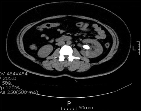
FIG. 1 Three regions of interest with a diameter of 2 mm were drawn on the stone in the axial plane of NCCT where the stone length was the longest. The mean number of HU calculated from the 3 regions represents the density of the stone. NCCT: non-contrast computed tomography, HU: Hounsfield unit.
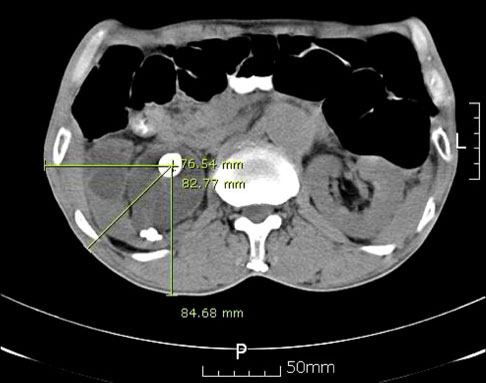
FIG. 2 Measurement of the skin-to-stone distance at 0°, 45°, and 90° on an axial scan of NCCT. NCCT: non-contrast computed tomography.
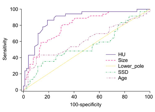
FIG. 3 Comparison of ROC curves to test the statistical significance of the difference between the area under different ROC curves. In pairwise comparison of all predictors for the outcome of shock wave lithotripsy, stone density and stone size was not different (AUC difference: 0.0465, p=0.388). ROC: receiver operator characteristic, AUC: area under the ROC curve.
Reference
-
1. Chaussy C, Brendel W, Schmiedt E. Extracorporeally induced destruction of kidney stones by shock waves. Lancet. 1980. 2:1265–1268.2. Lingeman JE, Newman D, Mertz JH, Mosbaugh PG, Steele RE, Kahnoski RJ, et al. Extracorporeal shock wave lithotripsy: the Methodist Hospital of Indiana experience. J Urol. 1986. 135:1134–1137.3. Ehreth JT, Drach GW, Arnett ML, Barnett RB, Govan D, Lingeman J, et al. Extracorporeal shock wave lithotripsy: multicenter study of kidney and upper ureter versus middle and lower ureter treatments. J Urol. 1994. 152:1379–1385.4. Cass AS. Comparison of first generation (Dornier HM3) and second generation (Medstone STS) lithotriptors: treatment results with 13,864 renal and ureteral calculi. J Urol. 1995. 153:588–592.5. Lingeman JE, Woods JR, Toth PD. Blood pressure changes following extracorporeal shock wave lithotripsy and other forms of treatment for nephrolithiasis. JAMA. 1990. 263:1789–1794.6. Kim JH, Moon YT. Predicting the therapeutic effect of extracorporeal shockwave lithotripsy by non-enhanced computed tomography in renal stones. Korean J Urol. 2008. 49:252–256.7. El-Nahas AR, El-Assmy AM, Mansour O, Sheir KZ. A prospective multivariate analysis of factors predicting stone disintegration by extracorporeal shock wave lithotripsy: the value of high-resolution noncontrast computed tomography. Eur Urol. 2007. 51:1688–1693.8. Dalla Palma L, Pozzi-Mucelli R, Stacul F. Present-day imaging of patients with renal colic. Eur Radiol. 2001. 11:4–17.9. Katz DS, Lane MJ, Sommer FG. Non-contrast spiral CT for patients with suspected renal colic. Eur Radiol. 1997. 7:680–685.10. Wang LJ, Ng CJ, Chen JC, Chiu TF, Wong YC. Diagnosis of acute flank pain caused by ureteral stones: value of combined direct and indirect signs on IVU and unenhanced helical CT. Eur Radiol. 2004. 14:1634–1640.11. Yilmaz S, Sindel T, Arslan G, Ozkaynak C, Karaali K, Kabaalioglu A, et al. Renal colic: comparison of spiral CT, US and IVU in the detection of ureteral calculi. Eur Radiol. 1998. 8:212–217.12. Arac M, Celik H, Oner AY, Gultekin S, Gumus T, Kosar S. Distinguishing pelvic phleboliths from distal ureteral calculi: thin-slice CT findings. Eur Radiol. 2005. 15:65–70.13. Preminger GM, Vieweg J, Leder RA, Nelson RC. Urolithiasis: detection and management with unenhanced spiral CT--a urologic perspective. Radiology. 1998. 207:308–309.14. Ahn SS, Lee SH, Kang I. The value of non-enhanced spiral CT in the diagnosis of suspected urolithiasis. Korean J Urol. 2002. 43:1008–1013.15. Gupta NP, Ansari MS, Kesarvani P, Kapoor A, Mukhopadhyay S. Role of computed tomography with no contrast medium enhancement in predicting the outcome of extracorporeal shock wave lithotripsy for urinary calculi. BJU Int. 2005. 95:1285–1288.16. Kim HS, Jang SW, Jeong YB, Kim YG, Kim JS. The usefulness of unenhanced helical computerized tomography in patients with urinary calculi. Korean J Urol. 2003. 44:796–800.17. Obek C, Onal B, Kantay K, Kalkan M, Yalcin V, Oner A, et al. The efficacy of extracorporeal shock wave lithotripsy for isolated lower pole calculi compared with isolated middle and upper caliceal calculi. J Urol. 2001. 166:2081–2084.18. Shiroyanagi Y, Yagisawa T, Nanri M, Kobayashi C, Toma H. Factors associated with failure of extracorporeal shock-wave lithotripsy for ureteral stones using Dornier lithotripter U/50. Int J Urol. 2002. 9:304–307.19. Abdel-Khalek M, Sheir K, Elsobky E, Showkey S, Kenawy M. Prognostic factors for extracorporeal shock-wave lithotripsy of ureteric stones--a multivariate analysis study. Scand J Urol Nephrol. 2003. 37:413–418.20. Abdel-Khalek M, Sheir KZ, Mokhtar AA, Eraky I, Kenawy M, Bazeed M. Prediction of success rate after extracorporeal shock-wave lithotripsy of renal stones--a multivariate analysis model. Scand J Urol Nephrol. 2004. 38:161–167.21. Srivastava A, Zaman W, Singh V, Mandhani A, Kumar A, Singh U. Efficacy of extracorporeal shock wave lithotripsy for solitary lower calyceal stone: a statistical model. BJU Int. 2004. 93:364–368.22. Lingeman JE, Coury TA, Newman DM, Kahnoski RJ, Mertz JH, Mosbaugh PG, et al. Comparison of results and morbidity of percutaneous nephrostolithotomy and extracorporeal shock wave lithotripsy. J Urol. 1987. 138:485–490.23. Psihramis KE, Jewett MA, Bombardier C, Caron D, Ryan M. Toronto Lithotripsy Associates. Lithostar extracorporeal shock wave lithotripsy: the first 1,000 patients. J Urol. 1992. 147:1006–1009.24. Daudon M, Donsimoni R, Hennequin C, Fellahi S, Le Moel G, Paris M, et al. Sex- and age-related composition of 10 617 calculi analyzed by infrared spectroscopy. Urol Res. 1995. 23:319–326.25. Pareek G, Armenakas NA, Fracchia JA. Hounsfield units on computerized tomography predict stone-free rates after extracorporeal shock wave lithotripsy. J Urol. 2003. 169:1679–1681.26. Pareek G, Armenakas NA, Panagopoulos G, Bruno JJ, Fracchia JA. Extracorporeal shock wave lithotripsy success based on body mass index and Hounsfield units. Urology. 2005. 65:33–36.27. Joseph P, Mandal AK, Singh SK, Mandal P, Sankhwar SN, Sharma SK. Computerized tomography attenuation value of renal calculus: can it predict successful fragmentation of the calculus by extracorporeal shock wave lithotripsy? A preliminary study. J Urol. 2002. 167:1968–1971.28. Wang LJ, Wong YC, Chuang CK, Chu SH, Chen CS, See LC, et al. Predictions of outcomes of renal stones after extracorporeal shock wave lithotripsy from stone characteristics determined by unenhanced helical computed tomography: a multivariate analysis. Eur Radiol. 2005. 15:2238–2243.29. Abe T, Akakura K, Kawaguchi M, Ueda T, Ichikawa T, Ito H, et al. Outcomes of shockwave lithotripsy for upper urinary-tract stones: a large-scale study at a single institution. J Endourol. 2005. 19:768–773.30. Pareek G, Hedican SP, Lee FT Jr, Nakada SY. Shock wave lithotripsy success determined by skin-to-stone distance on computed tomography. Urology. 2005. 66:941–944.
- Full Text Links
-
- Actions
-
Cited
- CITED
-
- Close
- Share
-
- Similar articles
-
- An Experience of Abdominal Aortic Pseudoaneurysm in a Patient Having Abdominal Aortic Aneurysmal Disease after Extracorporeal Shock Wave Lithotripsy
- Clinical Experience of Extracorporeal Shock Wave Lithotripsy for Urinary Calculi
- Extraction of Common Bile Duct Stones Refractory to Mechanical Lithotripsy Using Extracorporeal Shock Wave Lithotripsy
- Use of Renal Scan(DTPA) for Clinical Follow-up of Renal Function after Extracorporeal Shock Wave Lithotripsy of Renal Stones
- Effect of Extracorporeal Shock Wave Lithotripsy of Caliceal Stone according to the Location of the Stone
