Obstet Gynecol Sci.
2015 Nov;58(6):518-521. 10.5468/ogs.2015.58.6.518.
Two pregnancy cases of uterine scar dehiscence after laparoscopic myomectomy
- Affiliations
-
- 1Department of Obstetrics and Gynecology, Chungnam National University Hospital, Daejeon, Korea.
- 2Department of Obstetrics and Gynecology, Chungnam National University School of Medicine, Daejeon, Korea. minari73@cnuh.co.kr
- KMID: 2314070
- DOI: http://doi.org/10.5468/ogs.2015.58.6.518
Abstract
- Uterine scar dehiscence following laparoscopic myomectomy rarely occurs but can compromise both maternal and fetal well-being in subsequent pregnancy. We here present two cases of pregnancy complicated by preterm birth that resulted from uterine scar dehiscence following laparoscopic myomectomy. First case was a nulligravida who had scar dehiscence at 26 weeks of gestation after having a laparoscopic myomectomy 3 months prior to conception. Two weeks later, we observed her fetal leg protruding through the defect. The other case was a primigravida with a history of prior cesarean delivery, whose sonography revealed myomectomy scar dehiscence at 31 weeks of gestation. Within a few hours after observing, the patient complained of abdominal pain that was aggravating as fetal leg protruded through the defect. In both cases, babies were born by emergency cesarean section. Conservative management can be one of treatment options for myomectomy scar dehiscence in preterm pregnancy. However, clinicians should always be aware of the possibility of obstetric emergencies.
MeSH Terms
Figure
-
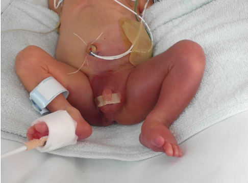
Fig. 1 The newborn's left leg shows bruise and edema.
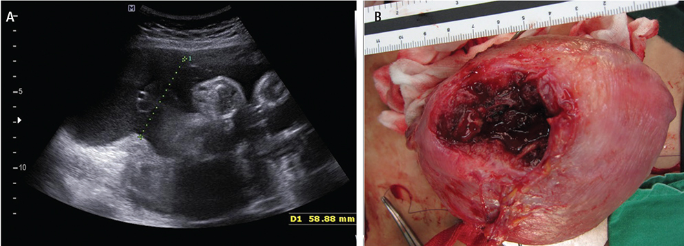
Fig. 2 Uterine wall dehiscence. (A) Ultrasonography showed the protrusion of the amniotic sac through the uterine defect. (B) Photograph of the uterine defect.
Reference
-
1. Hasbargen U, Summerer-Moustaki M, Hillemanns P, Scheidler J, Kimmig R, Hepp H. Uterine dehiscence in a nullipara, diagnosed by MRI, following use of unipolar electrocautery during laparoscopic myomectomy: case report. Hum Reprod. 2002; 17:2180–2182.2. Syam HH. Pregnancy outcomes following laparoscopic myomectomy. World J Laparosc Surg. 2008; 1:35–40.3. Roopnarinesingh S, Suratsingh J, Roopnarinesingh A. The obstetric outcome of patients with previous myomectomy or hysterotomy. West Indian Med J. 1985; 34:59–62.4. Banas T, Klimek M, Fugiel A, Skotniczny K. Spontaneous uterine rupture at 35 weeks' gestation, 3 years after laparoscopic myomectomy, without signs of fetal distress. J Obstet Gynaecol Res. 2005; 31:527–530.5. Dubuisson JB, Fauconnier A, Deffarges JV, Norgaard C, Kreiker G, Chapron C. Pregnancy outcome and deliveries following laparoscopic myomectomy. Hum Reprod. 2000; 15:869–873.6. Nezhat CH, Nezhat F, Roemisch M, Seidman DS, Tazuke SI, Nezhat CR. Pregnancy following laparoscopic myomectomy: preliminary results. Hum Reprod. 1999; 14:1219–1221.7. Paul PG, Koshy AK, Thomas T. Pregnancy outcomes following laparoscopic myomectomy and single-layer myometrial closure. Hum Reprod. 2006; 21:3278–3281.8. Veena P, Habeebullah S, Chaturvedula L. A review of 93 cases of ruptured uterus over a period of 2 years in a tertiary care hospital in South India. J Obstet Gynaecol. 2012; 32:260–263.9. Ronel D, Wiznitzer A, Sergienko R, Zlotnik A, Sheiner E. Trends, risk factors and pregnancy outcome in women with uterine rupture. Arch Gynecol Obstet. 2012; 285:317–321.10. Rabinowitz R, Samueloff A, Sapirstein E, Shen O. Expectant management of fetal arm extruding through a large uterine dehiscence following sonographic diagnosis at 27 weeks of gestation. Ultrasound Obstet Gynecol. 2006; 28:235–237.11. Rajab KE, Al-Ojaimi E, Sundari M. Spontaneous uterine rupture in the second trimester of pregnancy associated with red degeneration of fibroid. Bahrain Med Bull. 2006; 28:1–4.12. Oyelese Y, Tchabo JG, Chapin B, Nair A, Hanson P, McLaren R. Conservative management of uterine rupture diagnosed prenatally on the basis of sonography. J Ultrasound Med. 2003; 22:977–980.
- Full Text Links
-
- Actions
-
Cited
- CITED
-
- Close
- Share
-
- Similar articles
-
- Obstetric outcomes after uterine myomectomy: Laparoscopic versus laparotomic approach
- Repetitive Spontaneous Uterine Rupture in the First Trimester after Laparoscopic Myomectomy: A Case Report and Review of Literature
- Obstetric outcomes after laparoscopic myomectomy
- Comparsion of Laparoscopic with minilaparotomic myomectomy in uterine myoma
- Disseminated Peritoneal Leiomyomatosis Following Previous Laparoscopic Myomectomy with Morcellation
