Korean J Urol.
2008 Sep;49(9):855-859.
Leiomyosarcoma at the Ureterovesical Junction
- Affiliations
-
- 1Department of Urology, Wonkwang University School of Medicine, Institute of Wonkwang Medical Science, Iksan, Korea. sc.park@wonkwang.ac.kr
Abstract
- Non-urothelial neoplasms of the bladder account for less than 5% of all bladder tumors. Leiomyosarcoma is the most common malignant mesenchymal tumor that arises in the adult bladder. Leiomyosarcomas of the bladder are considered to be highly aggressive tumors. Most patients present at an advanced stage, with less than 30% of patients presenting with stage T1 disease. Surgical resection still remains the cornerstone of treatment with the status of the surgical margin being a strong predictor of the outcome. A 59-year-old man presented to our institution with urinary urgency, and cystoscopic examination revealed a huge submucosal mass on the lateral bladder wall. A radical cystoprostatectomy and ileal neobladder procedure was performed. The patient was diagnosed with a primary leiomyosarcoma of the bladder. After 1 year of follow-up, there has been no recurrence or metastasis.
Keyword
MeSH Terms
Figure
-
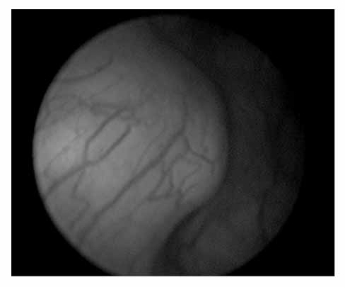
Fig. 1 Cystoscopic findings. There is an intramural mass on the right lateral wall of the bladder.
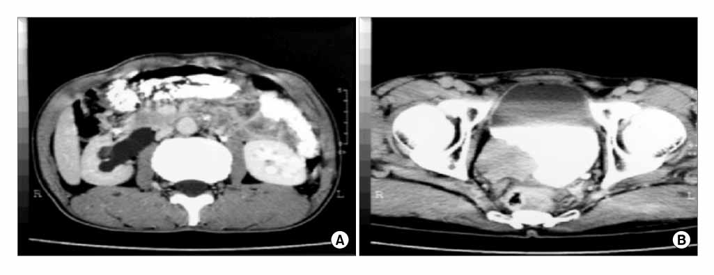
Fig. 2 Abdomino-pelvic CT scan. (A) Note the right-sided hydronephrosis with ureteral dilatation. (B) A 7×5cm low attenuation mass is seen at the right ureterovesical junction.
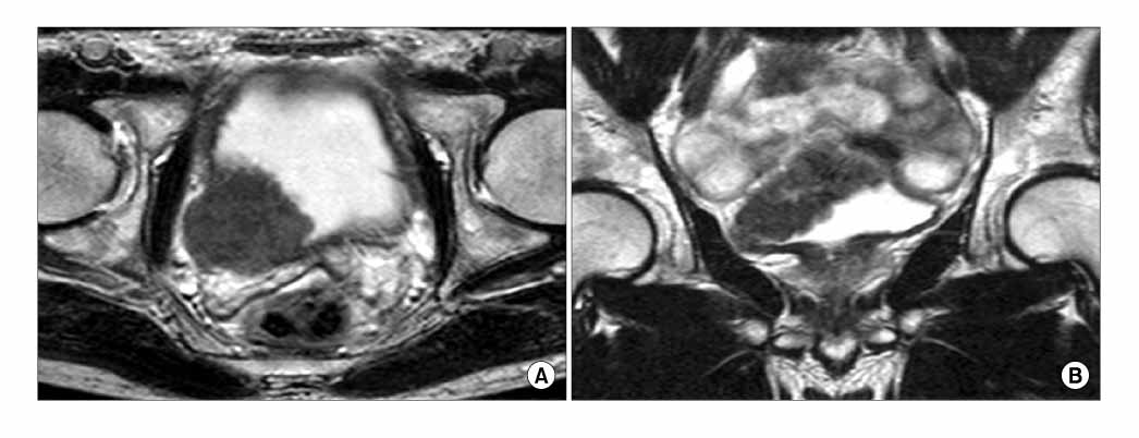
Fig. 3 Pelvic T2-weighted magnetic resonance imaging (MRI). A 7×5cm subtle high signal intensity mass is seen at the right bladder base and in the ureter.
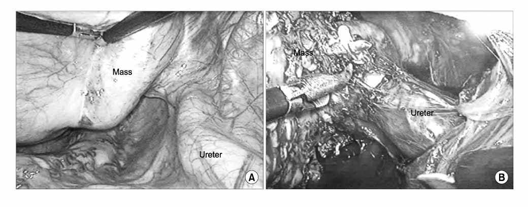
Fig. 4 Operative findings (laparoscopic view). (A) The mass protrudes from the right bladder wall. (B) Note the mass surrounding the ureter.
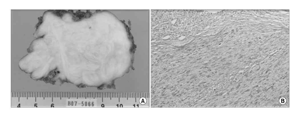
Fig. 5 (A) Gross findings: a 7×4.5cm sized whitish mass. (B) Microscopy shows the high cellularity and mild to moderate nuclear atypia (H&E, ×200).
Reference
-
1. Pedersen-Bjergaard J, Jonsson V, Pedersen M, Hou-Jensen K. Leiomyosarcoma of the urinary bladder after cyclophosphamide. J Clin Oncol. 1995. 13:532–533.2. Rosser CJ, Slaton JW, Izawa JI, Levy LB, Dinney CP. Clinical presentation and outcome of high-grade urinary bladder leiomyosarcoma in adults. Urology. 2003. 61:1151–1155.3. Russo P, Brady MS, Conlon K, Hajdu SI, Fair WR, Herr HW, et al. Adult urological sarcoma. J Urol. 1992. 147:1032–1036.4. Russell WO, Cohen J, Edmonson JH, Enzinger F, Hajdu SK, Heise H, et al. Staging system for soft tissue sarcoma. Semin Oncol. 1981. 8:156–159.5. Mills SE, Bova GS, Wick MR, Young RH. Leiomyosarcoma of the urinary bladder. A clinicopathologic and immunohistochemical study of 15 cases. Am J Surg Pathol. 1989. 13:480–489.6. Martin SA, Sears DL, Sebo TJ, Lohse CM, Cheville JC. Smooth muscle neoplasms of the urinary bladder: a clinicopathologic comparison of leiomyoma and leiomyosarcoma. Am J Surg Pathol. 2002. 26:292–300.7. Swartz DA, Johnson DE, Ayala AG, Watkins DL. Bladder leiomyosarcoma; a review of 10 cases with 5-year followup. J Urol. 1985. 133:200–202.8. De Berardinis E, Giulianelli R, Zarrelli G, De Santis C, Ginepri A, Gentile BC, et al. Leiomyosarcoma of urinary bladder: personal experience in 3 cases over a 10-year period. Arch Ital Urol Androl. 1997. 69:Suppl 1. 73–80.9. Strander H, Turesson I, Cavallin-Stahl E. A systematic overview of radiation therapy effects in soft tissue sarcomas. Acta Oncol. 2003. 42:516–531.10. Spiess PE, Kassouf W, Steinberg JR, Tuziak T, Hernandez M, Tibbs RF, et al. Review of the M.D. Anderson experience in the treatment of bladder sarcoma. Urol Oncol. 2007. 25:38–45.
- Full Text Links
-
- Actions
-
Cited
- CITED
-
- Close
- Share
-
- Similar articles
-
- A Case of Cecoureterocele with Contralateral Ureterovesical Junction Stricture
- Obstruction of the lower ureter by obliterated umbilical artyery: report of a case
- Cystic Dysplasia of the Testis Associated with Ipsilateral Hypoplastic Type of Renal Dysplasia and Ureterovesical Junction Obstruction
- Microsurgical Anatomy and the Function of the Ureterovesical Junction
- Urologic Complications of Renal Transplantation
