Korean J Urol.
2008 Dec;49(12):1094-1099.
Predicting Factors for Spontaneous Passage of Ureteral Calculi Based on Unenhanced Helical CT Findings
- Affiliations
-
- 1Department of Urology, Myongji Hospital, College of Medicine, Kwandong University, Goyang, Korea. urokdj@hotmail.com
Abstract
-
PURPOSE: We performed a prospective study to determine whether unenhanced helical computed tomography(UHCT) findings predict the spontaneous passage of ureteral calculi.
MATERIALS AND METHODS
Between April 2006 and June 2007, 175 patients with a single ureteral calculus <1cm in diameter were enrolled in this study, and a UHCT was performed. All of the patients were managed conservatively for 2 weeks. Patients without spontaneous passage of ureteral calculi within 2 weeks were treated by ureteroscopy or extracorporeal shock wave lithotripsy(ESWL). The secondary signs (hydronephrosis, perinephric edema, and the tissue rim sign) were graded on a scale of 0-3. We evaluated whether spontaneous passage of ureteral calculi was associated with stone diameter, location, Hounsfield units(HU), and the degree of secondary signs.
RESULTS
Ninety-two patients(52.6%) had spontaneous passage of ureteral calculi. The mean stone diameter was significantly smaller in the passage group than the non-passage group(4.28mm vs. 6.73mm, p=0.002). The rate of spontaneous passage was significantly higher involving distal ureteral calculi(66.1%) than proximal ureteral calculi(30.3%, p<0.001). The incidences of hydronephrosis and perinephric edema were significantly lower in the spontaneous passage group than the non-passage group(8.7% vs. 73.5% and 5.4% vs. 69.9%, respectively). The grades of hydronephrosis and perinephric edema were significantly lower in the spontaneous passage group than the non-passage group(p=0.001). Although there was a tendency toward increasing grades of hydronephrosis and perinephric edema with increasing stone size, the grades were more frequent and severe in the non-passage group in patients with similarly sized stones.
CONCLUSIONS
The degree of hydronephrosis and perinephric edema are useful ancillary signs in predicting the likelihood of spontaneous passage of ureteral calculi.
MeSH Terms
Figure
-
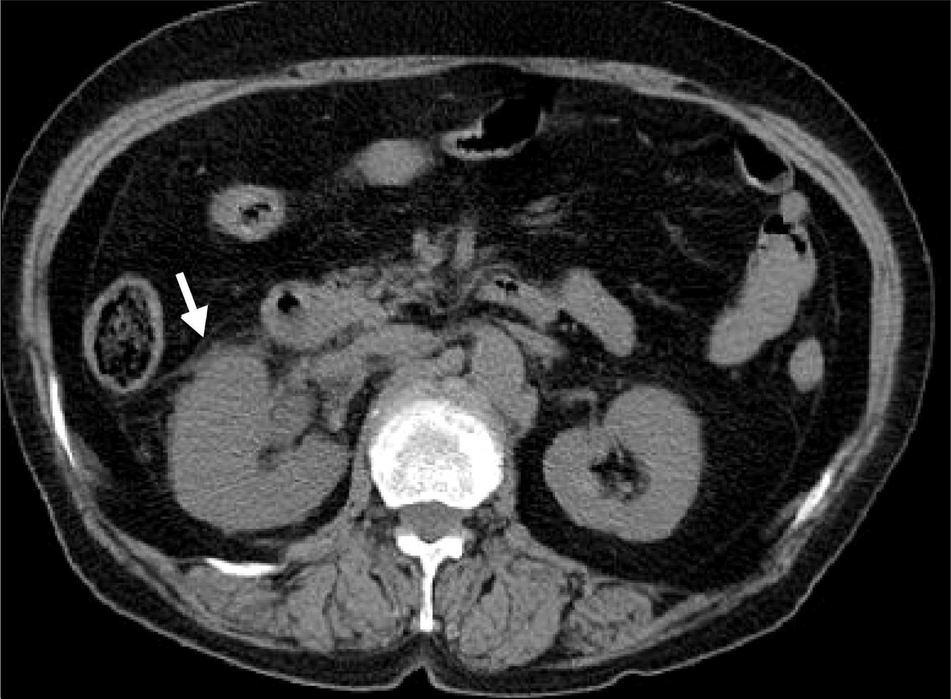
Fig. 1. Unenhanced helical CT revealed hydronephrosis, renal enlargement, and perinephric edema (white arrow) in a patient with acute right-sided flank pain.

Fig. 2. Axial unenhanced helical CT images showed ureteral stones and a tissue rim sign (white arrow).

Fig. 3. Incidence of secondary signs related to stone diameter.

Fig. 4. Mean grade of secondary signs related to stone diameter.
Reference
-
References
1. Katz DS, Lane MJ, Sommer FG. Unenhanced helical CT of ureteral stones: incidence of associated urinary tract findings. AJR Am J Roentgenol. 1996; 166:1319–22.
Article2. Coll DM, Varanelli MJ, Smith RC. Relationship of spontaneous passage of ureteral calculi to stone size and location as revealed by unenhanced helical CT. AJR Am J Roentgenol. 2002; 178:101–3.
Article3. Ege G, Akman H, Kuzucu K, Yildiz S. Acute ureterolithiasis: incidence of secondary signs on unenhanced helical CT and influence on patient management. Clin Radiol. 2003; 58:990–4.
Article4. Kwak SM, Moon YT, Kim SC. Spontaneous passage of ureteral stone by conservative treatment. Korean J Urol. 1993; 34:308–12.5. Sandegard E. Prognosis of stone in the ureter. Acta Chir Scand. 1956; 219(Suppl):1–67.6. Porpiglia F, Destefanis P, Fiori C, Fontana D. Effectiveness of nifedipine and deflazacort in the management of distal ureteral stones. Urology. 2000; 56:579–82.7. Fox M, Pyrah LN, Raper FP. Management of ureteric stone: a review of 292 cases. Br J Urol. 1965; 37:660–70.8. Smith RC, Rosenfield AT, Choe KA, Essenmacher KR, Verga M, Glickman MG, et al. Acute flank pain: comparison of non-contrast-enhanced CT and intravenous urography. Radiology. 1995; 194:789–94.
Article9. Fielding JR, Steele G, Fox LA, Heller H, Loughlin KR. Spiral computerized tomography in the evaluation of acute flank pain: a replacement for excretory urography. J Urol. 1997; 157:2071–3.
Article10. Smith RC, Dalrymple NC, Neitlich J. Noncontrast helical CT in the evaluation acute flank pain. Abdom Imaging. 1998; 23:10–6.11. Winfield AC, Gerlock AJ Jr, Shaff MI. Perirenal cobwebs: a CT sign of renal vein thrombosis. J Comput Assist Tomogr. 1981; 5:705–8.
Article12. Takahashi N, Kawashima A, Ernst RD, Boridy IC, Goldman SM, Benson GS, et al. Ureterolithiasis: can clinical outcome be predicted with unenhanced helical CT? Radioloogy. 1998; 208:97–102.
Article13. Kawashima A, Sandler CM, Bofidy IC, Takahashi N, Benson GS, Goldman SM. Unenhanced helical CT of ureterolithiasis: value of the tissue rim sign. AJR Am J Roentgenol. 1997; 168:997–1000.
Article14. Lumerman J, Gershbaum MD, Hines J, Nardi P, Beuchert P, Katz DS. Unenhanced helical computed tomography for the evaluation of suspected renal colic in the adolescent population: a pilot study. Urology. 2001; 57:342–6.
Article15. Zarse CA, McAteer JA, Tann M, Sommer AJ, Kim SC, Paterson RF, et al. Helical computed tomography accurately reports urinary stone composition using attenuation values: in vitro verification using high resolution micro computed tomography calibrat-ed to fourie transform infrared microspectroscopy. Urology. 2004; 63:828–33.
- Full Text Links
-
- Actions
-
Cited
- CITED
-
- Close
- Share
-
- Similar articles
-
- Comparision of Unenhanced Helical Computerized Tomography and Intravenous Urography in the Radiologic Evaluation of Acute Flank Pain
- The Usefulness of Unenhanced Helical Computerized Tomography in Patients with Urinary Calculi
- Spontaneous passage of ureteral stone by conservative treatment
- Unenhanced Spiral CT in Acute Ureteral Colic: A Replacement for Excretory Urography?
- Ureteral calculi: treatment options under advanced technology
