Does C5 or C6 Radiculopathy Affect the Signal Intensity of the Brachial Plexus on Magnetic Resonance Neurography?
- Affiliations
-
- 1Department of Rehabilitation Medicine, Keimyung University School of Medicine, Daegu, Korea. ri-pheonix@hanmail.net
- 2Pain Research Center, Keimyung University School of Medicine, Daegu, Korea.
- 3Institute for Medical Science, Keimyung University School of Medicine, Daegu, Korea.
- 4Department of Neurosurgery, Keimyung University School of Medicine, Daegu, Korea.
- 5Department of Orthopedic Surgery, Keimyung University School of Medicine, Daegu, Korea.
- KMID: 2309938
- DOI: http://doi.org/10.5535/arm.2016.40.2.362
Abstract
- Patients with C5 or C6 radiculopathy complain of shoulder area pain or shoulder girdle weakness. Typical idiopathic neuralgic amyotrophy (INA) is also characterized by severe shoulder pain, followed by paresis of shoulder girdle muscles. Recent studies have demonstrated that magnetic resonance neurography (MRN) of the brachial plexus and magnetic resonance imaging (MRI) of the shoulder in patients with INA show high signal intensity (HSI) or thickening of the brachial plexus and changes in intramuscular denervation of the shoulder girdle. We evaluated the value of brachial plexus MRN and shoulder MRI in four patients with typical C5 or C6 radiculopathy. HSI of the brachial plexus was noted in all patients and intramuscular changes were observed in two patients who had symptoms over 4 weeks. Our results suggest that HSI or thickening of the brachial plexus and changes in intramuscular denervation of the shoulder girdle on MRN and MRI may not be specific for INA.
MeSH Terms
Figure
-
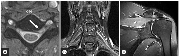
Fig. 1 Images of case 1. (A) C5-6 intervertebral disc herniation (arrow) compressing the left C6 root. (B) High signal intensity (HSI) in the C6 root (arrowheads) on coronal T2 short-tau inversion recovery (STIR) image. (C) HSI in the supraspinatus (asterisk) on coronal T2 STIR image.
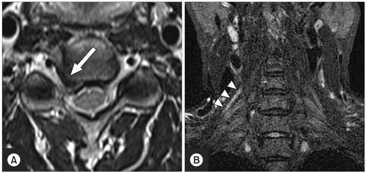
Fig. 2 Images of case 2. (A) C4-5 intervertebral disc herniation (arrow) compressing the right C5 root. (B) High signal intensity in the C5 root (arrowheads) on coronal T2 short-tau inversion recovery image.
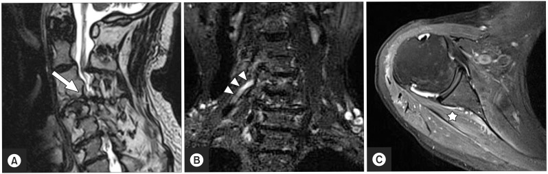
Fig. 3 Images of case 3. (A) Right C4-5 foraminal stenosis (arrow) compressing the C5 root. (B) High signal intensity (HSI) in the C5 root (arrowheads) on coronal T2 short-tau inversion recovery image. (C) HSI in the infraspinatus (asterisk) on axial T2-weighed image.

Fig. 4 Images of case 4. (A) Left C5-6 foraminal stenosis (arrow) compressing the C6 root. (B) Thickening of the C6 root (arrowheads) on coronal T2 short-tau inversion recovery image.
Reference
-
1. Woods BI, Hilibrand AS. Cervical radiculopathy: epidemiology, etiology, diagnosis, and treatment. J Spinal Disord Tech. 2015; 28:E251–E259. PMID: 25985461.2. van Alfen N, van Engelen BG. The clinical spectrum of neuralgic amyotrophy in 246 cases. Brain. 2006; 129(Pt 2):438–450. PMID: 16371410.
Article3. Duman I, Guvenc I, Kalyon TA. Neuralgic amyotrophy, diagnosed with magnetic resonance neurography in acute stage: a case report and review of the literature. Neurologist. 2007; 13:219–221. PMID: 17622915.4. Park MS, Kim DH, Sung DH. Magnetic resonance neurographic findings in classic idiopathic neuralgic amyotrophy in subacute stage: a report of four cases. Ann Rehabil Med. 2014; 38:286–291. PMID: 24855627.
Article5. Lee YS, Choi ES, Song CJ. Symptomatic nerve root changes on contrast-enhanced MR imaging after surgery for lumbar disk herniation. AJNR Am J Neuroradiol. 2009; 30:1062–1067. PMID: 19213822.
Article6. van Alfen N. The neuralgic amyotrophy consultation. J Neurol. 2007; 254:695–704. PMID: 17446996.
Article
- Full Text Links
-
- Actions
-
Cited
- CITED
-
- Close
- Share
-
- Similar articles
-
- Magnetic Resonance Neurography Findings in Idiopathic Neuralgic Amyotrophy
- An Updated Review of Magnetic Resonance Neurography for Plexus Imaging
- Quantification of the Nerve Fiber of the Terminal Branches of the Typical Brachial Plexus
- Anatomical Studies of the Upper and Lower Subscapular Nerves
- Magnetic Resonance Neurography with Short Tau Inversion Recovery Sequences for Cervical Radiculopathy
