Korean J Phys Anthropol.
2014 Dec;27(4):219-223. 10.11637/kjpa.2014.27.4.219.
Anatomical Studies of the Upper and Lower Subscapular Nerves
- Affiliations
-
- 1Department of Anatomy, Catholic Kwandong University College of Medicine, Korea. kslee@cku.ac.kr
- KMID: 1982459
- DOI: http://doi.org/10.11637/kjpa.2014.27.4.219
Abstract
- This study aimed to determine the number of branches of the upper and lower subscapular nerves and to classify the spinal nerve compositions of each branch. Twenty sides of the brachial plexus extracted from Korean adult cadavers were used in this study. The upper subscapular nerve was composed of three branches in 16 sides (80.0%), composed of four branches in 4 sides (20.0%). The lower subscapular nerve arose from posterior cord with one branch, innervating the subscapularis and the teres major muscles. In case of the upper subscapular nerve, the first branch was comprised of C5 in 40.0%, C5 and C6 in 60.0%, the second branch was composed of C5 in 15.0%, C5 and C6 in 85.0%, the third branch was arisen from C5 and C6 in 75.0%, C6 in 25.0% and the forth branch appeared in four sided was derived from C6. The nerve branch innervating the subscapularis muscle was composed of C6 in 55.0%, C5 and C6 in 40.0%, C6 and C7 in 5.0%. The nerve branch innervating the teres major muscle was composed of C6 and C7 in 65.0%, C5, C6 and C7 in 25.0%, and C6 in 10.0%. The injury was often due to an accidental damage or lesion of the subscapular nerve, these anatomical results may be helpful to predict the involving area of the lesion.
Figure
-
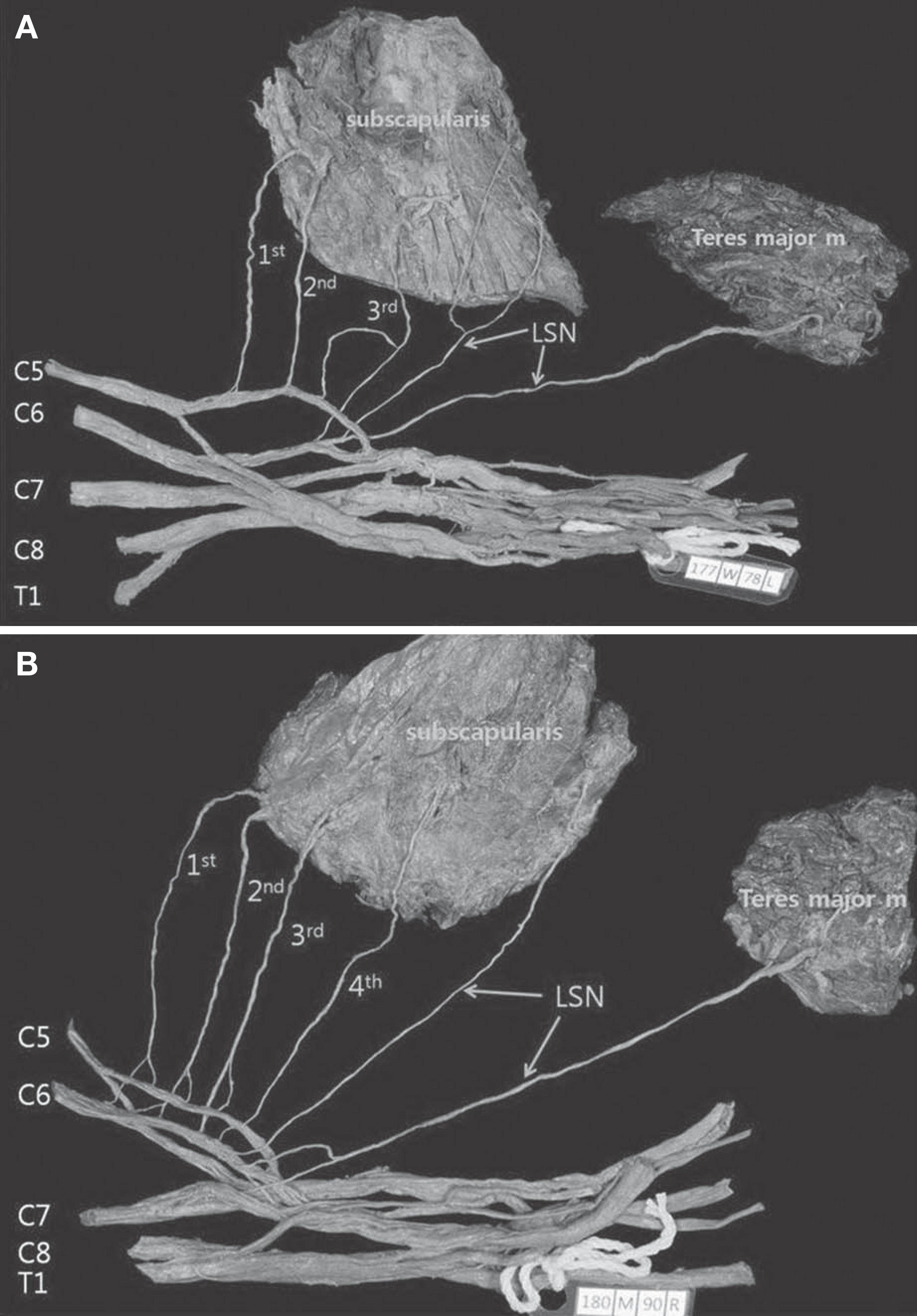
Fig. 1. Photographs show the number of the branches of the upper subscapular nerve (USN). A: The USN was comprised of the three branches(1st, 2nd, 3rd). B: The USN was comprised of the four branches(1st, 2nd, 3rd, 4th). LSN: lower subscapular nerve
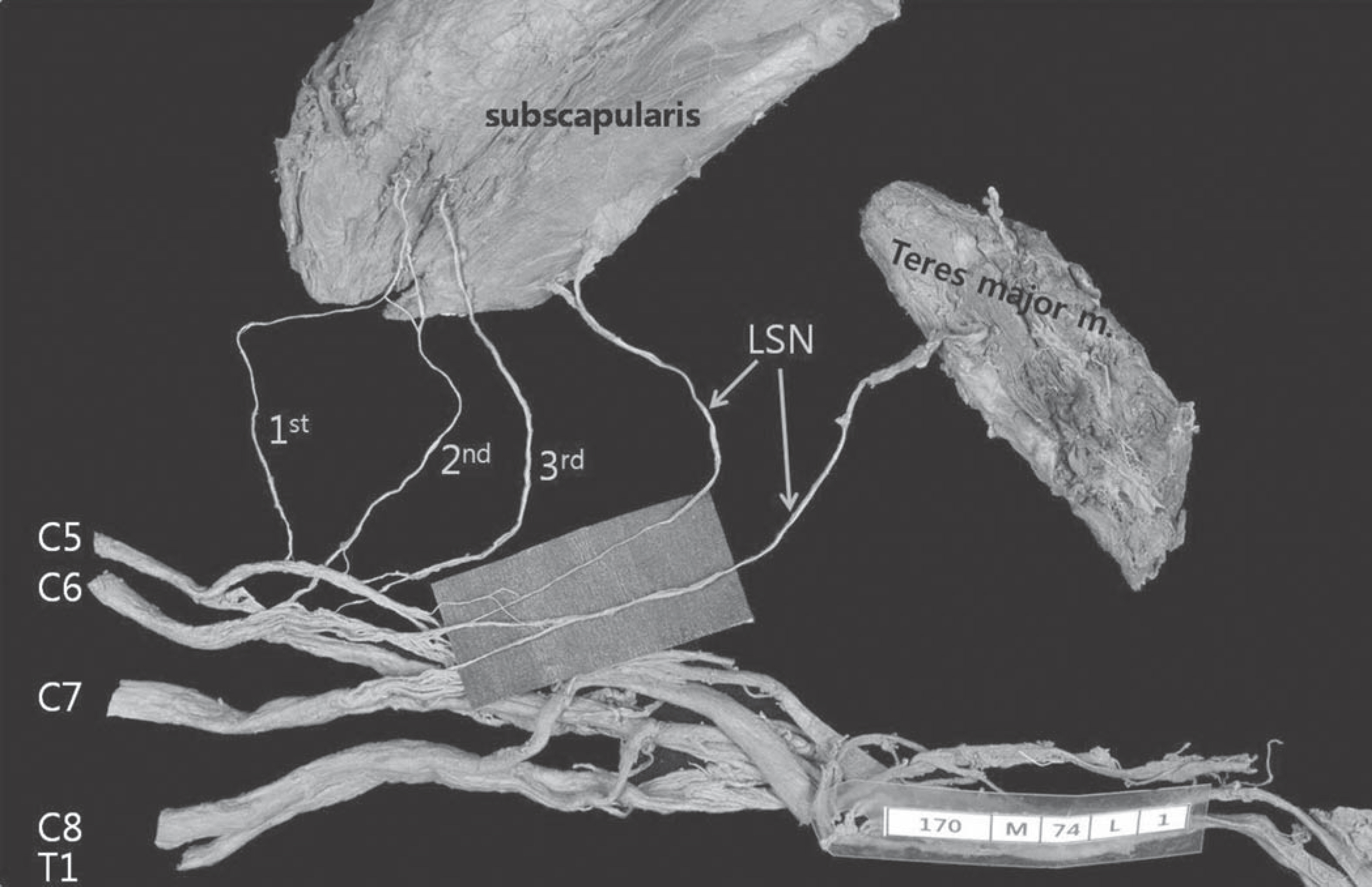
Fig. 2. A photograph shows the spinal nerve compositions of the branch of lower subscapular nerve (LSN). The C5 and C6 were distributed to the subscapularis muscle and C5, C6 and C7 were distributed to the teres major muscle.
Reference
-
References
1. Chung IH, Oh CS, Han SH, Kim HJ. Human Anatomy. 5th ed.Hyunmoon;2011. p. 81–97. Korean.2. Bergman RA, Thompson SA, Afifi AK, Saadeh FA. Compendium of human anatomic variation. Baltimore: Urban & Schwarzenberg;1988. p. 140.3. Woodburne RT, Burkel WE. Essentials of Human Anatomy. 9th ed.New York: Oxford University Press;1994. p. p. 121.4. Moore KL, Dalley AF. Clinically oriented anatomy. 5th ed.Philadelphia: Lippincott Williams&Wilkins;2006. p. 773–7.5. Muthoka JM, Sinkeet SR, Shahbal SH, Matakwa LC, Og-eng'o JA. Variations in branching of the posterior cord of brachial plexus in a Kenyan population. J Brachial Plex Peripher Nerve Inj. 2011; 6:1.
Article6. Tubbs RS, Loukas M, Shahid K, Judge T, Pinyard J, et al. Anatomy and quantitation of the subscapular nerves. Clin Anat. 2007; 20:656–9.
Article7. Saleh DB, Callear J, McConnell P, Kay SP. The anatomy of the subscapular nerves: a new nomenclature. J Plast Reconstr Aesthet Surg. 2012; 65:1072–5.
Article8. Ballesteros LE, Ramirez LM. Variations of the origin of collateral branches emerging from the posterior aspect of the brachial plexus. J Brachial Plex Peripher Nerve Inj. 2007; 2:14.
Article
- Full Text Links
-
- Actions
-
Cited
- CITED
-
- Close
- Share
-
- Similar articles
-
- Usefullness of Chimeric Flaps Based on the Subscapular Vascular System
- Quantification of the Nerve Fiber of the Terminal Branches of the Typical Brachial Plexus
- Alternative prosthetic vascular access creation using subscapular artery as inflow to prevent dialysis access related steal syndrome
- Branching pattern and morphometry of the axillary nerve
- Variations in Branching Patterns of the Anterior Circumflex Humeral Artery
