Human nasociliary nerve with special reference to its unique parasympathetic cutaneous innervation
- Affiliations
-
- 1Division of Ophthalmology, Iwamizawa Municipal Hospital, Iwamizawa, Japan.
- 2Department of Anatomy, Tokyo Dental College, Chiba, Japan. yamamotomasahito@tdc.ac.jp
- 3Department of Neurology, Wonkwang University School of Medicine and Hospital, Institute of Wonkwang Medical Science, Iksan, Korea.
- 4Division of Physical Therapy, Ongoul Rehabilitation Hospital, Jeonju, Korea.
- 5Division of Internal Medicine, Iwamizawa Kojin-kai Hospital, Iwamizawa, Japan.
- KMID: 2308920
- DOI: http://doi.org/10.5115/acb.2016.49.2.132
Abstract
- The frontal nerve is characterized by its great content of sympathetic nerve fibers in contrast to cutaneous branches of the maxillary and mandibular nerves. However, we needed to add information about composite fibers of cutaneous branches of the nasociliary nerve. Using cadaveric specimens from 20 donated cadavers (mean age, 85), we performed immunohistochemistry of tyrosine hydroxylase (TH), neuronal nitric oxide synthase (nNOS), and vasoactive intestinal polypeptide (VIP). The nasocilliary nerve contained abundant nNOS-positive fibers in contrast to few TH- and VIP-positive fibers. The short ciliary nerves also contained nNOS-positive fibers, but TH-positive fibers were more numerous than nNOS-positive ones. Parasympathetic innervation to the sweat gland is well known, but the original nerve course seemed not to be demonstrated yet. The present study may be the first report on a skin nerve containing abundant nNOS-positive fibers. The unique parasympathetic contents in the nasocilliary nerve seemed to supply the forehead sweat glands as well as glands in the eyelid and nasal epithelium.
Keyword
MeSH Terms
Figure
-
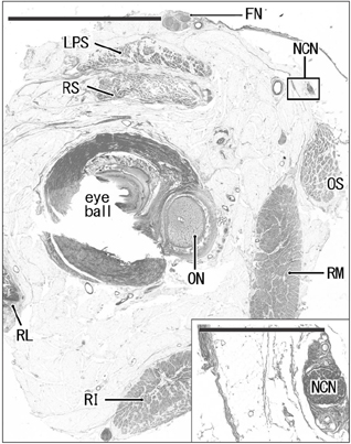
Fig. 1 Topographical anatomy of the nasociliary and frontal nerves (a 78-year-old woman). Frontal section of the right eye. Hematoxylin and eosin staining. In the present study, the nasociliary nerve (NCN) was identified in a large frontal section including the posterior pole of the eye ball (scale bar=10 mm) and we traced the nerve anteriorly toward the skin. An insert at the lower angle is a higher magnification view of a square including the nasociliary nerve (scale bar=1 mm). FN, frontal nerve; LPS, levator palpebrae superioris muscle; ON, optic nerve; OS, obliquus superior muscle; RI, rectus inferior muscle; RL, rectus lateralis muscle; RM, rectus medialis muscle; RS, rectus superior muscle.
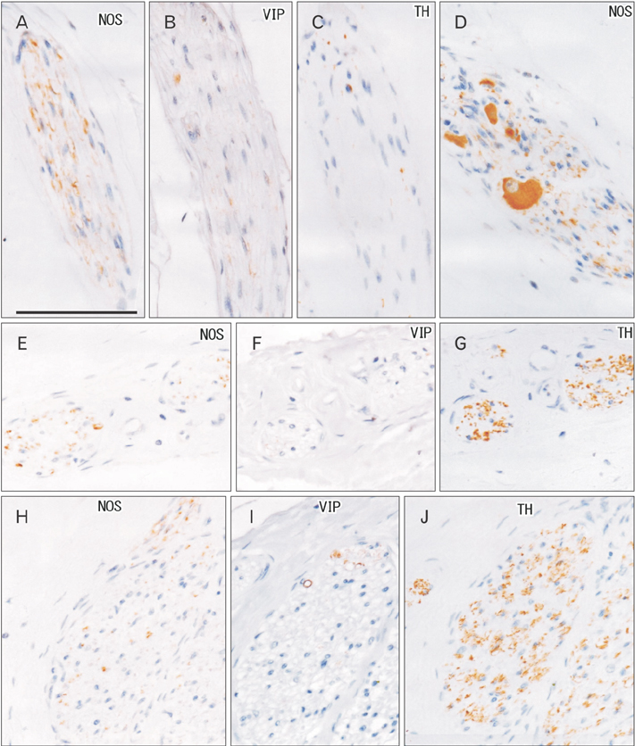
Fig. 2 Immunohistochemistr y of nerves (a 78-year-old woman). The specimen same as in Fig. 1. Frontal sections almost 2 mm anterior to Fig. 1. (A–D) The nasocilliary nerve (A–C, near sections). (E–G) A ciliary nerve (near sections). (H–J) The frontal nerve (near sections). (A, D, E, H) Immunohistochemistry of neuronal nitric oxide synthase (NOS). (B, F, I) Immunohistochemistry of vasoactive intestinal polypeptide (VIP). (C, G, J) Immunohistochemistry of tyrosine hydroxylase (TH). The nasociliary nerve contains abundant NOS-positive nerve fibers, while TH-positive fibers are dominant in the frontal nerve. A branch of the nasociliary nerve contains NOS-positive ganglion cells (D). All panels are prepared at the same magnification. Scale bar=0.1 mm.
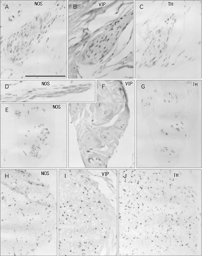
Fig. 3 Immunohistochemistry of nerves (an 89-year-old man). Frontal sections. (A–D) The nasocilliary nerve (A–C, near sections). (E–G) A ciliary nerve (near sections). (H–J) The frontal nerve (near sections). (A, D, E, H) Immunohistochemistry of neuronal nitric oxide synthase (NOS). (B, F, I) Immunohistochemistry of vasoactive intestinal polypeptide (VIP). (C, G, J) Immunohistochemistry of tyrosine hydroxylase (TH). The nasocilliary nerve contain abundant NOS-positive nerve fibers, while TH-positive fibers are dominant in the frontal nerve. (D) A branch of the nasocilliary nerve. All panels are prepared at the same magnification. Scale bar=0.1 mm.
Reference
-
1. Matsubayashi T, Cho KH, Jang HS, Murakami G, Yamamoto M, Abe SI. Significant differences in sympathetic nerve fiber density among the facial skin nerves: a histologic study using human cadaveric specimens. Anat Rec (Hoboken). 2016; 04. 12. [Epub]. DOI: 10.1002/ar.23347.2. Baljet B, van der Werf F, Otto AJ. Autonomic pathways in the orbit of the human fetus and the rhesus monkey. Doc Ophthalmol. 1989; 72:247–264.3. Oikawa S, Kawagishi K, Yokouchi K, Fukushima N, Moriizumi T. Immunohistochemical determination of the sympathetic pathway in the orbit via the cranial nerves in humans. J Neurosurg. 2004; 101:1037–1044.4. Thakker MM, Huang J, Possin DE, Ahmadi AJ, Mudumbai R, Orcutt JC, Tarbet KJ, Sires BS. Human orbital sympathetic nerve pathways. Ophthal Plast Reconstr Surg. 2008; 24:360–366.5. Rusu MC, Pop F. The anatomy of the sympathetic pathway through the pterygopalatine fossa in humans. Ann Anat. 2010; 192:17–22.6. Kiyokawa H, Katori Y, Cho KH, Murakami G, Kawase T, Cho BH. Reconsideration of the autonomic cranial ganglia: an immunohistochemical study of mid-term human fetuses. Anat Rec (Hoboken). 2012; 295:141–149.7. Nordin M. Sympathetic discharges in the human supraorbital nerve and their relation to sudo- and vasomotor responses. J Physiol. 1990; 423:241–255.8. Cramer MN, Bain AR, Jay O. Local sweating on the forehead, but not forearm, is influenced by aerobic fitness independently of heat balance requirements during exercise. Exp Physiol. 2012; 97:572–582.9. Yuzuriha S, Matsuo K, Ishigaki Y, Kikuchi N, Kawagishi K, Moriizumi T. Efferent and afferent innervations of Mueller's muscle related to involuntary contraction of the levator muscle: important for avoiding injury during eyelid surgery. Br J Plast Surg. 2005; 58:42–52.10. Hinata N, Hieda K, Sasaki H, Murakami G, Abe S, Matsubara A, Miyake H, Fujisawa M. Topohistology of sympathetic and parasympathetic nerve fibers in branches of the pelvic plexus: an immunohistochemical study using donated elderly cadavers. Anat Cell Biol. 2014; 47:55–65.11. Kellogg DL Jr, Zhao JL, Wu Y. Neuronal nitric oxide synthase control mechanisms in the cutaneous vasculature of humans in vivo. J Physiol. 2008; 586:847–857.12. Ikeyama K, Denda S, Tsutsumi M, Denda M. Neuronal nitric oxide synthase in epidermis is involved in cutaneous circulatory response to mechanical stimulation. J Invest Dermatol. 2010; 130:1158–1166.13. Del Pozzi AT, Carter SJ, Collins AB, Hodges GJ. The regional differences in the contribution of nitric oxide synthase to skin blood flow at forearm and lower leg sites in response to local skin warming. Microvasc Res. 2013; 90:106–111.14. Wong BJ. Sensory nerves and nitric oxide contribute to reflex cutaneous vasodilation in humans. Am J Physiol Regul Integr Comp Physiol. 2013; 304:R651–R656.15. Ibba-Manneschi L, Niissalo S, Milia AF, Allanore Y, Del Rosso A, Pacini A, Manetti M, Toscano A, Cipriani P, Liakouli V, Giacomelli R, Kahan A, Konttinen YT, Matucci-Cerinic M. Variations of neuronal nitric oxide synthase in systemic sclerosis skin. Arthritis Rheum. 2006; 54:202–213.16. Schulze E, Witt M, Fink T, Hofer A, Funk RH. Immunohistochemical detection of human skin nerve fibers. Acta Histochem. 1997; 99:301–309.17. Balogh B, Auterith A, Behrus R, Maier S, Vesely M, Piza-Katzer H. The sympathetic axons of the nerves of the hand. Handchir Mikrochir Plast Chir. 2002; 34:369–373.18. Marx SC, Kumar P, Dhalapathy S, Marx CA, D'Souza AS. Distribution of sympathetic fiber areas of radial nerve in the forearm: an immunohistochemical study in cadavers. Surg Radiol Anat. 2010; 32:865–871.19. Kinugasa Y, Arakawa T, Murakami G, Fujimiya M, Sugihara K. Nerve supply to the internal anal sphincter differs from that to the distal rectum: an immunohistochemical study of cadavers. Int J Colorectal Dis. 2014; 29:429–436.20. Nakao T, Cho KH, Yamamoto M, Yamane S, Murakami G, Ide Y, Abe S. Site-dependent difference in the density of sympathetic nerve fibers in muscle-innervating nerves: a histologic study using human cadavers. Eur J Anat. 2012; 16:33–42.21. Hosaka F, Katori Y, Kawase T, Fujimiya M, Ohguro H. Site-dependent differences in density of sympathetic nerve fibers in muscle-innervating nerves of the human head and neck. Anat Sci Int. 2014; 89:101–111.22. Hieda K, Cho KH, Arakawa T, Fujimiya M, Murakami G, Matsubara A. Nerves in the intersphincteric space of the human anal canal with special reference to their continuation to the enteric nerve plexus of the rectum. Clin Anat. 2013; 26:843–854.23. Guidry G, Landis SC. Absence of cholinergic sympathetic innervation from limb muscle vasculature in rats and mice. Auton Neurosci. 2000; 82:97–108.24. Hardebo JE, Suzuki N, Ekblad E, Owman C. Vasoactive intestinal polypeptide and acetylcholine coexist with neuropeptide Y, dopamine-beta-hydroxylase, tyrosine hydroxylase, substance P or calcitonin gene-related peptide in neuronal subpopulations in cranial parasympathetic ganglia of rat. Cell Tissue Res. 1992; 267:291–300.25. Wang N, Gibbons CH, Freeman R. Novel immunohistochemical techniques using discrete signal amplification systems for human cutaneous peripheral nerve fiber imaging. J Histochem Cytochem. 2011; 59:382–390.
- Full Text Links
-
- Actions
-
Cited
- CITED
-
- Close
- Share
-
- Similar articles
-
- Nasociliary Nerve Radiofrequency Thermocoagulation for Trigeminal Neuralgia: A case report
- Learning about Dermatome Maps and Innervation of Peripheral Cutaneous Nerves Using OHP film-Overlapping
- Unusual bilateral sensory innervation of the dorsum of hand by lateral antebrachial cutaneous nerve: a case report
- Frey' s Syndrome in a Child without Definite Causes
- Nerve-sparing radical hysterectomy: time for a new standard of care for cervical cancer?
