Anomalous Origin of the Left Circumflex Coronary Artery From the First Diagonal Branch Presented as Acute Myocardial Infarction
- Affiliations
-
- 1Division of Cardiology, Gimcheon Jeil Hospital, Gimcheon, Korea.
- 2Division of Cardiology, Daegu Catholic University Medical Center, Daegu, Korea. mdleeys@cu.ac.kr
- 3Pohang St. Mary Hospital, Pohang, Korea.
- 4Division of Cardiology, Dongguk University Medical Center, Gyeongju, Korea.
- 5Division of Radiology, Daegu Catholic University Medical Center, Daegu, Korea.
- KMID: 2297921
- DOI: http://doi.org/10.4070/kcj.2011.41.10.612
Abstract
- Coronary artery anomalies are diagnosed in 0.6 to 1.5% of patients who undergo coronary angiography (CAG). They may present with life threatening conditions but are generally asymptomatic. Recognition and adequate visualization of the anomaly is essential for correct management of the condition. However, in some cases the exact orifice and course of an anomalous coronary vessel cannot be selectively identified by CAG. In this report, a 54-year-old man was admitted to the hospital with acute inferior myocardial infarction and had an anomalous origin of the left circumflex coronary artery (LCX) from the first diagonal branch (D1). In CAG, the right CAG showed no significant stenosis and fortunately we found an anomalous origin of the LCX from the D1. The course of LCX was precisely established by 64-slice multi-detector computed tomography.
Keyword
MeSH Terms
Figure
-
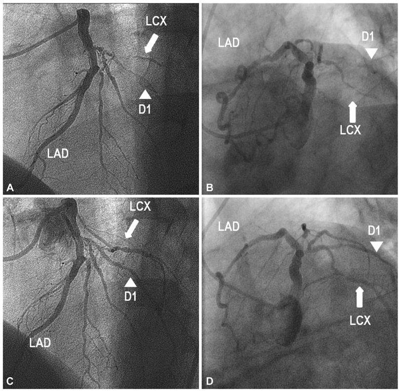
Fig. 1 Coronary angiographic finding. A and B: coronary angiography shows the small artery (arrow) with TIMI flow grade 1 originating from the D1 (arrowhead). C and D: after percutaneous coronary intervention for the small artery, the coronary angiography reveals an anomalous origin of the LCX from the D1. A and C: left anterior oblique cranial view. B and D: left anterior oblique caudal views. TIMI: thrombolysis in myocardial infarction, LAD: left anterior descending coronary artery, LCX: left circumflex coronary artery, D1: first diagonal branch.

Fig. 2 64-slice multi-detector computed tomography demonstrates an anomalous origin of the LCX coursed to the left atrioventricular (AV) groove from the D1. LAD: left anterior descending coronary artery, LCX: left circumflex coronary artery, D1: first diagonal branch, LA: left atrium, LV: left ventricle.
Reference
-
1. Yamanaka O, Hobbs RE. Coronary artery anomalies in 126,595 patients undergoing coronary arteriography. Cathet Cardiovasc Diagn. 1990. 21:28–40.2. Gavrielatos G, Letsas KP, Pappas LK, Antonellis I, Kardaras F. Anomalous origin of the entire coronary system with separate ostia within the right sinus of valsalva: a rare congenital anomaly and a review of the literature. Cardiology. 2007. 107:209–212.3. Aydinlar A, Cicek D, Senturk T, et al. Primary congenital anomalies of the coronary arteries: a coronary arteriographic study in Western Turkey. Int Heart J. 2005. 46:97–103.4. Wilkins CE, Betancourt B, Mathur VS, et al. Coronary artery anomalies: review of over 10,000 patients from the Clayton Cardiovascular Laboratories. Tex Heart Inst J. 1988. 15:166–173.5. Lee YS, Lee JB, Kim KS. Anomalous origin of the left circumflex coronary artery from the right sinus of valsalva identified by imaging with multidetector computed tomography. Korean Circ J. 2006. 36:823–825.6. Korosoglou G, Ringwald G, Giannitsis E, Katus HA. Anomalous origin of the left circumflex coronary artery from the pulmonary artery: a very rare congenital anomaly in an adult patient diagnosed by cardiovascular magnetic resonance. J Cardiovasc Magn Reson. 2008. 10:4.7. Post JC, van Rossum AC, Bronzwaer JG, et al. Magnetic resonance angiography of anomalous coronary arteries: a new gold standard for delineating the proximal course. Circulation. 1995. 92:3163–3171.
- Full Text Links
-
- Actions
-
Cited
- CITED
-
- Close
- Share
-
- Similar articles
-
- Correlation between Abnormal Q Wave in Leads I or aVL and First Diagonal Branch Stenosis in Patients with Acute Myocardial Infarction
- Anomalous Origin of the Left Coronary Artery from the Right Sinus of Valsalva, which Presented as Acute Myocardial Infarction
- Congenital Absence of Left Circumflex Coronary Artery: Circumflex Artery Extended from Right Coronary Artery
- Anomalous Separate Origin of Left Anterior Descending Coronary Artery: Presented as Acute Anterior Myocardial Infarction
- Role of Transesophageal Echocardiography in Identifying Anomalous Origin and Course of Coronary Arteries
