Transcatheter Treatment of Atrial Septal Defect Presenting with Platypnea-Orthodeoxia Syndrome
- Affiliations
-
- 1Division of Pediatirc Cardiology, Congenital Heart Disease Center, Severance Cardiovascular Hospital, Department of Pediatrics, Yonsei University College of Medicine, Seoul, Korea. cjy0122@yuhs.ac
- 2Department of Pediatrics, Gangnam Severance Hospital, Yonsei University College of Medicine, Seoul, Korea.
- KMID: 2297886
- DOI: http://doi.org/10.4070/kcj.2015.45.2.169
Abstract
- A 29-year-old woman was referred to our institute for symptomatic hypoxemia. Her dyspnea was aggravated while sitting or standing and relieved while in supine position. She did not have any pulmonary disease. Transthoracic echocardiography and heart computed tomography revealed an underestimated small atrial septal defect (ASD) with a left-to-right shunt. A cardiac catheterization was performed to evaluate pulmonary hypertension. It revealed a normal pulmonary artery pressure and a large ASD with bidirectional shunt during Valsalva maneuver by intracardiac echocardiogram. Her arterial oxygen saturation decreased from 93% while supine to 79% while at a 15degrees sitting position. Thus, the patient was diagnosed with platypnea-orthodeoxia syndrome. The ASD was successfully closed with Amplatzer(R) (St. Jude Medical) septal occluder and both platypnea and orthodeoxia were resolved immediately after the procedure.
MeSH Terms
Figure
-
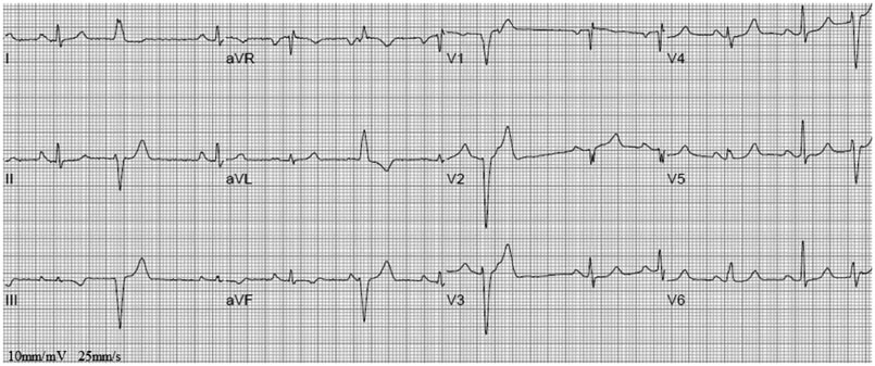
Fig. 1 Twelve-lead electrocardiography. The electrocardiography shows premature ventricular complexes and RSr' pattern in V 1.
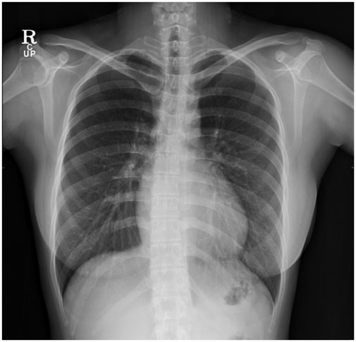
Fig. 2 Chest X-ray on admission. A chest X-ray radiography showed no pulmonary lesion and no cardiomegaly (cardio-thoracic ratio was 48%).

Fig. 3 Intracardiac echocardiography. A: an ICE showed an underestimated large ASD. B: the Doppler imaging showed a left-to-right shunt through an ASD. C: however, a bidirectional shunt occurred with Valsalva maneuver. ICE: intracardiac echocardiography, ASD: atrial septal defect, LA: left atrium, RA: right atrium.

Fig. 4 A large ASD was treated with Amplatzer® (St. Jude Medical) septal occluder. A large ASD was measured by the balloon occlusion method (A) and successfully treated with 36-mm sized Amplatzer® (St. Jude Medical) septal occluder (B). Her symptoms were resolved immediately after the procedure. ASD: atrial septal defect.
Reference
-
1. Burchell HB, Helmholz HF Jr, Wood EH. Reflex orthostatic dyspnea associated with pulmonary hypertension. Am J Physiol. 1949; 159:563–564.2. Seward JB, Hayes DL, Smith HC, et al. Platypnea-orthodeoxia: clinical profile, diagnostic workup, management, and report of seven cases. Mayo Clin Proc. 1984; 59:221–231.3. Cheng TO. Platypnea-orthodeoxia syndrome: etiology, differential diagnosis, and management. Catheter Cardiovasc Interv. 1999; 47:64–66.4. Robin ED, Laman D, Horn BR, Theodore J. Platypnea related to orthodeoxia caused by true vascular lung shunts. N Engl J Med. 1976; 294:941–943.5. Robin ED, McCauley RF. An analysis of platypnea-orthodeoxia syndrome including a "new" therapeutic approach. Chest. 1997; 112:1449–1451.6. Rodrigues P, Palma P, Sousa-Pereira L. Platypnea-orthodeoxia syndrome in review: defining a new disease? Cardiology. 2012; 123:15–23.7. van Gaal WJ, Joseph M, Jones E, Matalanis G, Horrigan M. Platypnea-orthodeoxia associated with a fenestrated atrial septal aneurysm: case report. Cardiovasc Ultrasound. 2005; 3:28.8. Natalie AA, Nichols L, Bump GM. Platypnea-orthodeoxia, an uncommon presentation of patent foramen ovale. Am J Med Sci. 2010; 339:78–80.9. Hashimoto M, Okawa Y, Baba H, Nishimura Y, Aoki M. Platypnea-orthodeoxia syndrome combined with constrictive pericarditis after coronary artery bypass surgery. J Thorac Cardiovasc Surg. 2006; 132:1225–1226.10. Adolph EA, Lacy WO, Hermoni YI, Wexler LF, Javaheri S. Reversible orthodeoxia and platypnea due to right-to-left intracardiac shunting related to pericardial effusion. Ann Intern Med. 1992; 116:138–139.11. Rao PS, Palacios IF, Bach RG, Bitar SR, Sideris EB. Platypnea-orthodeoxia: management by transcatheter buttoned device implantation. Catheter Cardiovasc Interv. 2001; 54:77–82.12. Hirai N, Fukunaga T, Kawano H, et al. Platypnea - orthodeoxia syndrome with atrial septal defect. Circ J. 2003; 67:172–175.13. Tobis MJ, Azarbal B. Does patent foramen ovale promote cryptogenic stroke and migraine headache? Tex Heart Inst J. 2005; 32:362–365.14. Mortelmans K, Post M, Thijs V, Herroelen L, Budts W. The influence of percutaneous atrial septal defect closure on the occurrence of migraine. Eur Heart J. 2005; 26:1533–1537.15. Luermans JG, Post MC, Temmerman F, et al. Is a predominant left-to-right shunt associated with migraine?: A prospective atrial septal defect closure study. Catheter Cardiovasc Interv. 2009; 74:1078–1084.16. Delgado G, Inglessis I, Martin-Herrero F, et al. Management of platypnea-orthodeoxia syndrome by transcatheter closure of atrial communication: hemodynamic characteristics, clinical and echocardiographic outcome. J Invasive Cardiol. 2004; 16:578–582.
- Full Text Links
-
- Actions
-
Cited
- CITED
-
- Close
- Share
-
- Similar articles
-
- Platypnea-Orthodeoxia Syndrome Two Decades after Definitive Surgical Repair of Pulmonary Atresia with Intact Ventricular Septum
- Transcatheter Closure of Secundum Atrial Septal Defect with the Amplatzer Septal Occluder
- Comprehensive understanding of atrial septal defects by imaging studies for successful transcatheter closure
- Outcome of Transcatheter Closure of Oval Shaped Atrial Septal Defect with Amplatzer Septal Occluder
- Procedural, Early and Long-Term Outcomes after Transcatheter Atrial Septal Defects Closure: Comparison between Large and Very Large Atrial Septal Defect Groups
