Korean J Urol.
2006 Mar;47(3):344-346. 10.4111/kju.2006.47.3.344.
Calcifying Fibrous Pseudotumor of the Spermatic Cord
- Affiliations
-
- 1Department of Urology, College of Medicine, Hanyang University, Seoul, Korea. hychoi1@yahoo.co.kr
- 2Department of Pathology, College of Medicine, Hanyang University, Seoul, Korea.
- KMID: 2294186
- DOI: http://doi.org/10.4111/kju.2006.47.3.344
Abstract
- Calcifying fibrous pseudotumor is an uncommon benign soft tissue lesion that is newly recognized in the literature. It predominately occurs in young adults and it is mainly located in the extremities, trunk and neck. The pathologic features of calcifying fibrous pseudotumor are characterized by a hyalinized fibrous stroma, psammomatous or dystrophic calcifications and chronic inflammatory cells. The pathogenesis of calcifying fibrous pseudotumor is uncertain. Total excision of the lesion is curative and recurrence is rare. We report here on a case of calcifying fibrous pseudotumor of the spermatic cord that was discovered as a palpable inguinal mass in a 24-year-old man.
Keyword
Figure
-
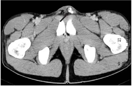
Fig. 1 Abdominal computed tomography shows a calcified mass along the left spermatic cord that measures about 1.6cm in diameter.
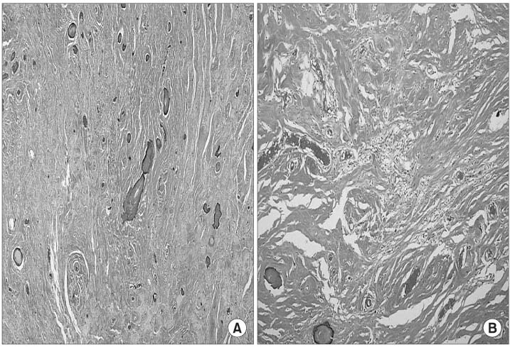
Fig. 2 Microscopic appearance shows areas of diffuse and extensive calcification with numerous psammomatous calcifications and other areas that are densely collagenized (A), and there are patchy infiltrates of plasma cells and lymphocytes in focal areas of the mass (B) (hematoxylin and eosin; ×100).
Reference
-
1. Pomplun S, Goldstraw P, Davies SE, Burke MM, Nicholson AG. Calcifying fibrous pseudotumor arising within an inflammatory pseudotumor: evidence of progression from one lesion to the other? Histopathology. 2000. 37:380–382.2. Fukunaga N, Kikuchi Y, Endo Y, Ushigome S. Calcifying fibrous pseudotumor. Pathol Int. 1997. 47:60–63.3. Nascimento AF, Ruiz R, Hornick JL, Fletcher CD. Calcifying fibrous 'pseudotumor': clinicopathologic study of 15 cases and analysis of its relationship to inflammatory myofibroblastic tumor. Int J Surg Pathol. 2002. 10:189–196.4. Zamecnik M, Michal M, Boudova L, Sulc M. CD34 expression in calcifying fibrous pseudotumours. Histopathology. 2000. 36:183–184.5. Youm MH, Shin SH, Kim SH, Cho HJ, Kim DY, Yoon GS. Calcifying fibrous pseudotumor in adrenal gland. Korean J Urol. 2003. 44:1298–1301.6. Rosenthal NS, Abdul-Karim FW. Childhood fibrous tumor with psammoma bodies. Clinicopathologic features in two cases. Arch Pathol Lab Med. 1988. 112:798–800.7. Fetsch JF, Montgomery EA, Meis JM. Calcifying fibrous pseudotumor. Am J Surg Pathol. 1993. 17:502–508.8. Van Dorpe J, Ectors N, Geboes K, D'Hoore A, Sciot R. Is calcifying fibrous pseudotumor a late sclerosing stage of inflammatory myofibroblastic tumor? Am J Surg Pathol. 1999. 23:329–335.9. Sigel JE, Smith TA, Reith JD, Goldblum JR. Immunohistochemical analysis of anaplastic lymphoma kinase expression in deep soft tissue calcifying fibrous pseudotumor: evidence of a late sclerosing stage of inflammatory myofibroblastic tumor? Ann Diagn Pathol. 2001. 5:10–14.10. Maeda T, Hirose T, Furuya K, Kameoka K. Calcifying fibrous pseudotumor: an ultrastructural study. Ultrastruct Pathol. 1999. 23:189–192.
- Full Text Links
-
- Actions
-
Cited
- CITED
-
- Close
- Share
-
- Similar articles
-
- Calcifying Fibrous Pseudotumor of the Retroperitoneum: A Case Report
- Calcifying fibrous pseudotumor of mediastinum--a case report
- Calcifying Fibrous Pseudotumor in Adrenal Gland
- Calcifying Fibrous Pseudotumor of the Anterior Mediastinum
- A Case of Fibrous Pseudotumor Originating from the Testicular Albuginea in the Children
