Osteochondral Lesion of Humeral Head Associated with Shoulder Internal Impingement: Report of Three Cases
- Affiliations
-
- 1Department of Orthopedic Surgery, Soonchunhyang University College of Medicine, Cheonan, Korea. cyclopia@naver.com
- KMID: 2288691
- DOI: http://doi.org/10.5763/kjsm.2014.32.1.59
Abstract
- Internal impingement syndrome is characterized by the posterior shoulder pain when the arm is abducted and external rotated, and articular partial rotator cuff tear with posterosuperior labral fraying in throwing athletes. Osteochondral lesion of humeral head as an associated lesion is reported in some cases but, not considered to be a main origin of the symptoms. We found the similar features of osteochondral lesion on humeral head in three cases of internal impingement syndrome irrespective of conservative treatment for over three months and report good results obtained from arthroscopic debridement and microfracturing for these lesions with a review of the literatures.
Keyword
MeSH Terms
Figure
-
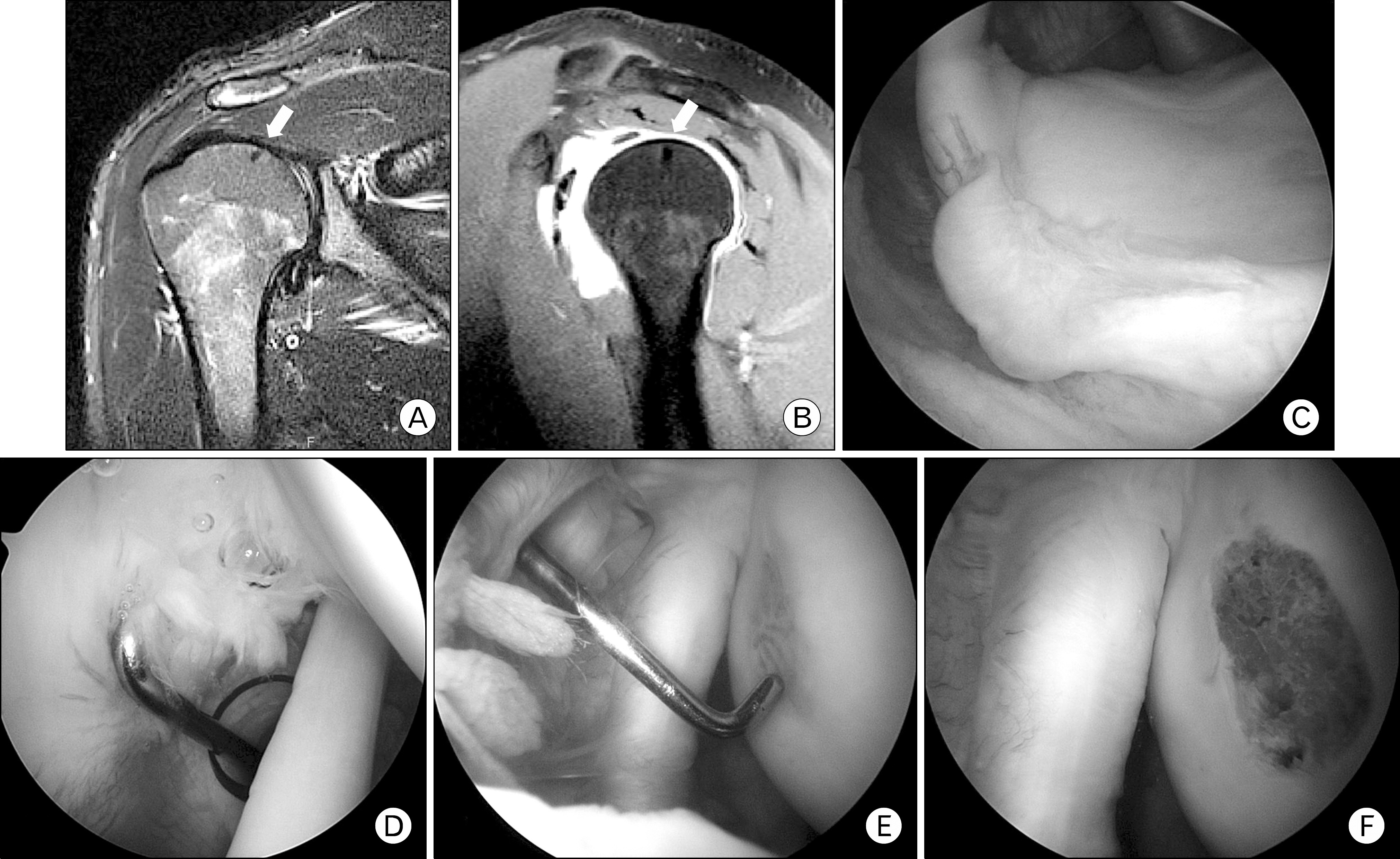
Fig. 1. (A, B) Coronal and sagittal magnetic resonance images show no significant osteochondral lesion of humeral head, but suspected lesion with low signal intensity as subchondral lesion (white arrow). (C) Arthroscopic finding shows posterosuperior labral fraying. (D) Arthroscopic finding shows intraarticular partial tear of supraspinatus tendon. But humeral head may seem like intact in 30o arthroscope viewing. (E) Osteochondral lesion of humeral head is confirmed by switching 70o arthroscope. (F) Arthroscopic debridement and microfracturing are performed.
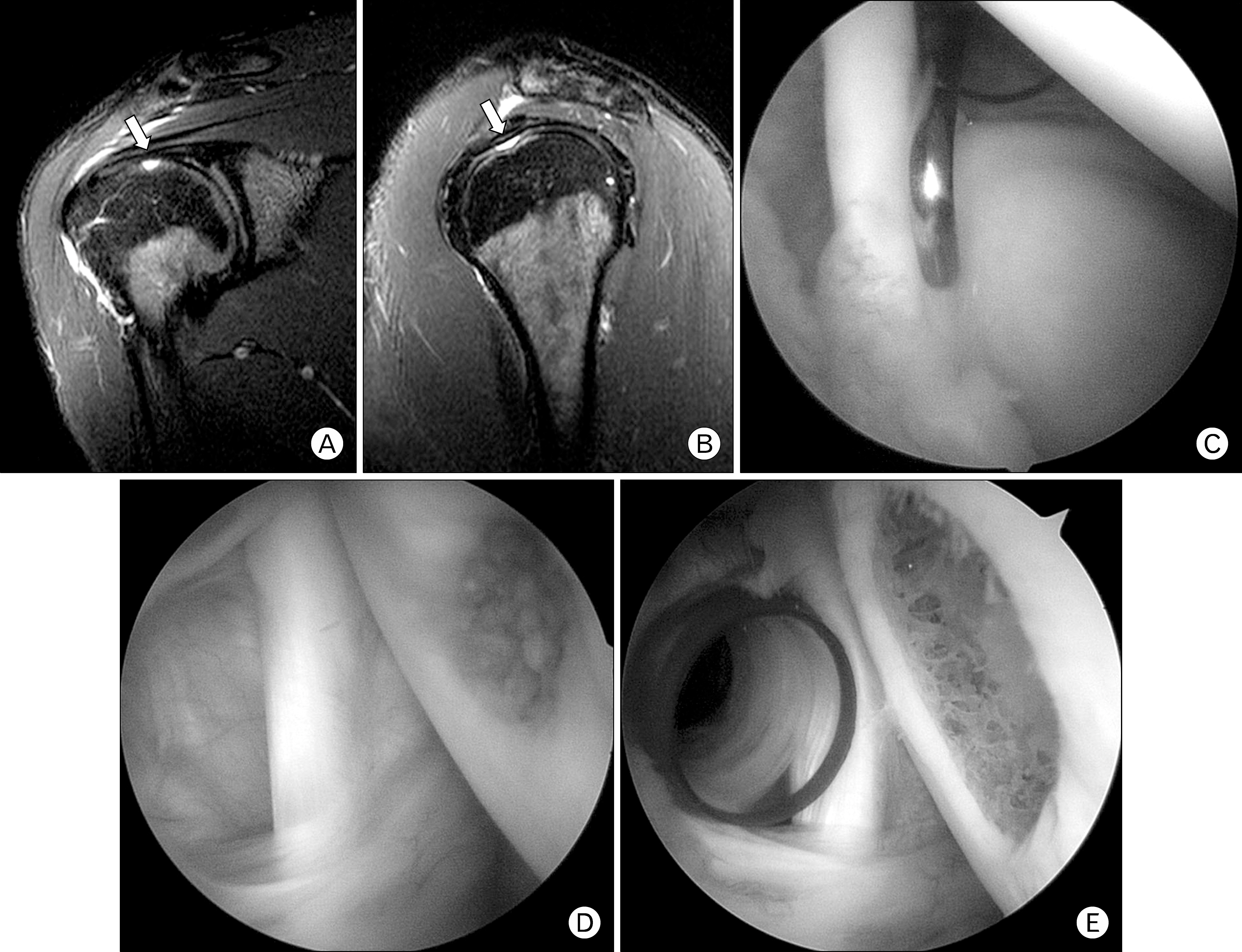
Fig. 2. (A, B) Magnetic resonance images show a focal chondral defect on the superior aspect of humeral head with high signal intensity on T2-weighted images (white arrow). (C) Arthroscopic findings show posterosuperior labral fraying. (D) Arthroscopic finding shows osteochondral lesion of humeral head. (E) Arthroscopic debridement is perfomed.
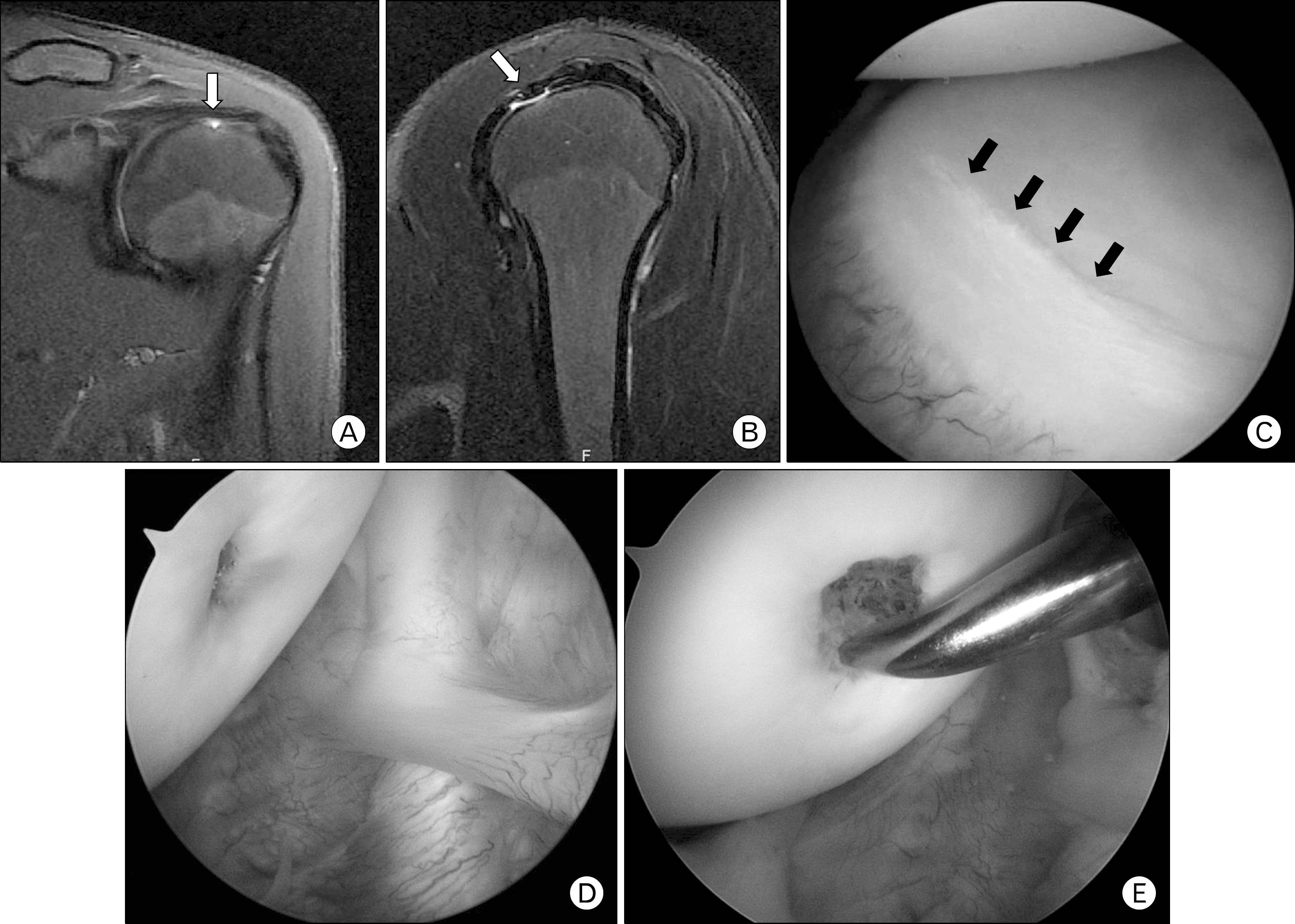
Fig. 3. (A, B) T2-weighted magnetic resonance images show high signal intensity lesion suspected as a chondral lesion (white arrow). (C) Arthroscopic finding shows stable scratches on the posterior glenoid labrum (black arrows). (D) Arthroscopic findings show focal chondral lesion of humeral head. (E) Debridement and microfracturing are perfomed arthroscopically.
Reference
-
1. Walch G, Boileau P, Noel E, Donell ST. Impingement of the deep surface of the supraspinatus tendon on the posterosuperior glenoid rim: An arthroscopic study. J Shoulder Elbow Surg. 1992; 1:238–45.
Article2. Davidson PA, Elattrache NS, Jobe CM, Jobe FW. Rotator cuff and posterior-superior glenoid labrum injury associated with increased glenohumeral motion: a new site of impingement. J Shoulder Elbow Surg. 1995; 4:384–90.
Article3. Heyworth BE, Williams RJ 3rd. Internal impingement of the shoulder. Am J Sports Med. 2009; 37:1024–37.
Article4. Jobe CM. Posterior superior glenoid impingement: expanded spectrum. Arthroscopy. 1995; 11:530–6.
Article5. Halbrecht JL, Tirman P, Atkin D. Internal impingement of the shoulder: comparison of findings between the throwing and nonthrowing shoulders of college baseball players. Arthroscopy. 1999; 15:253–8.
Article6. Burkhart SS, Morgan CD, Kibler WB. The disabled throwing shoulder: spectrum of pathology Part I: pathoanatomy and biomechanics. Arthroscopy. 2003; 19:404–20.
Article7. Meister K, Buckley B, Batts J. The posterior impingement sign: diagnosis of rotator cuff and posterior labral tears secondary to internal impingement in overhand athletes. Am J Orthop (Belle Mead NJ). 2004; 33:412–5.8. Mithofer K, Fealy S, Altchek DW. Arthroscopic treatment of internal impingement of the shoulder. Tech Shoulder Elbow Surg. 2004; 5:66–75.9. Paley KJ, Jobe FW, Pink MM, Kvitne RS, ElAttrache NS. Arthroscopic findings in the overhand throwing athlete: evidence for posterior internal impingement of the rotator cuff. Arthroscopy. 2000; 16:35–40.
Article
- Full Text Links
-
- Actions
-
Cited
- CITED
-
- Close
- Share
-
- Similar articles
-
- Chronic locked anterior shoulder dislocation with impaction of the humeral head onto the coracoid: a case report
- Dislocation of the Shoulder with Ipsilateral Humeral Shaft Fracture: A Case Report
- Osteonecrosis of the Humeral Head after Cerebral Angiography
- Ultrasonography in the Shoulder Impingement Syndrome
- Rapidly Progressive Osteonecrosis of the Humeral Head after Arthroscopic Bankart and Rotator Cuff Repair in a 66-Year Old Woman: A Case Report
