The Clinical Utility of Automated Breast Volume Scanner: A Pilot Study of 139 Cases
- Affiliations
-
- 1Department of Surgery, Chonbuk National University Medical School, Jeonju, Korea. yhj0903@jbnu.ac.kr
- 2Research Institute of Clinical Medicine, Chonbuk National University-Biomedical Research Institute, Chonbuk National University Hospital, Jeonju, Korea.
- 3Department of Radiology, Chonbuk National University Medical School, Jeonju, Korea.
- KMID: 2286385
- DOI: http://doi.org/10.4048/jbc.2013.16.3.329
Abstract
- PURPOSE
The aim of this study is to evaluate the clinical utility of automated breast volume scanner (ABVS) for detecting and diagnosing the breast lesions.
METHODS
From December 2010 to January 2012, bilateral whole breast examinations were performed with ABVS for 139 women. Based on the Breast Imaging Reporting and Data System (BI-RADS) categories, the breast lesions were evaluated on coronal multiplanar reconstruction images using the ABVS workstation. Then, the imaging results were compared with those on conventional handheld ultrasound (HHUS) images. Histological diagnoses were performed on BI-RADS category 4 and 5 lesions.
RESULTS
A total of 453 lesions were detected by ABVS. On the HHUS, 33 new lesions were detected but 69 lesions were not detected. BI-RADS category 2 and 3 matched to those on ABVS at 73.5% (61/83) and 85.4% (276/323). In 47 lesions of BI-RADS category 4 or 5, there was an exact match to those on ABVS. In addition, 47 lesions were classified as BI-RADS category 4 and 5, for which an ultrasound-guided core needle biopsy was performed. The malignant lesions of BI-RADS category 4 and 5 showed the following: 2/27 (7.4%) in 4A, 4/5 (80%) in 4B, 2/2 (100%) in 4C, and 13/13 (100%) in 5. The ABVS showed 21 true positives and a positive predictive value of 44.7% (21/47).
CONCLUSION
There was considerable agreement in the assessment of the breast lesions by ABVS and HHUS. The ABVS had advantages of high diagnostic accuracy, examiner-independence, multislice visualization of the whole breast and less time-consuming. Our results indicate that ABVS might be a useful modality in diagnosing breast lesions.
MeSH Terms
Figure
-

Figure 1 Automated breast volume scanner (ABVS) system (ACUSON S2000™; Siemens, Berlin, Germany). (A) The overall view of ABVS system. (B) A 14L5BV transducer (14 MHz, 15.4 cm) that has been designed for the ABVS.
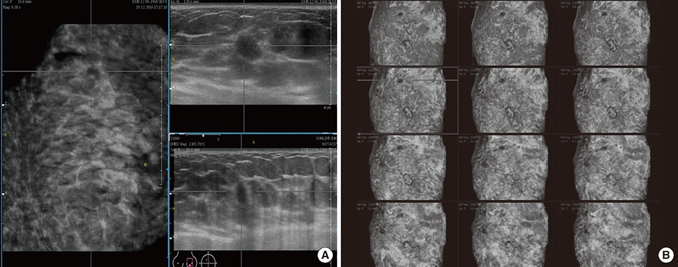
Figure 2 Multiplanar reconstruction of the volume data displayed on automated breast volume scanner. (A) Three on one view images that are provided following a multiplanar reconstruction using a workstation. Coronal view (left), longitudinal (right, upper), and transverse views (right, lower) are synchronously visualized on the screen. (B) A multislice view that has been designed to create serial sections of the images of overall breast in a similar manner to the computed tomography.
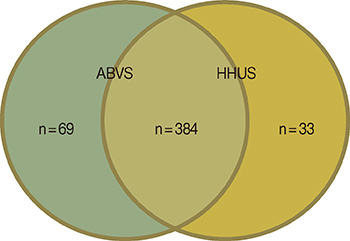
Figure 3 Breast mass on automated breast volume scanner (ABVS) and handheld ultrasound (HHUS). Additionally, 67 lesions were detected on the ABVS and 33 lesions were detected on the HHUS.

Figure 4 Comparison of automated breast volume scanner (ABVS) and handheld ultrasound (HHUS). A comparison of the images which were interpreted as the lesions of the Breast Imaging Reporting and Data System (BI-RADS) category 4B in HHUS (A) and ABVS (B). This lesion was confirmed to invasive ductal carcinoma by histologic examination. Additionally, a comparison of the images which were interpreted as the lesions of the BI-RADS category 3 in HHUS (C) and ABVS (D).
Reference
-
1. Humphrey LL, Helfand M, Chan BK, Woolf SH. Breast cancer screening: a summary of the evidence for the U.S. Preventive Services Task Force. Ann Intern Med. 2002; 137(5 Part 1):347–360.
Article2. Kolb TM, Lichy J, Newhouse JH. Comparison of the performance of screening mammography, physical examination, and breast US and evaluation of factors that influence them: an analysis of 27,825 patient evaluations. Radiology. 2002; 225:165–175.
Article3. Dempsey PJ. The history of breast ultrasound. J Ultrasound Med. 2004; 23:887–894.
Article4. Berg WA. Tailored supplemental screening for breast cancer: what now and what next? AJR Am J Roentgenol. 2009; 192:390–399.
Article5. Singh S, Tourassi GD, Baker JA, Samei E, Lo JY. Automated breast mass detection in 3D reconstructed tomosynthesis volumes: a featureless approach. Med Phys. 2008; 35:3626–3636.
Article6. Kotsianos-Hermle D, Hiltawsky KM, Wirth S, Fischer T, Friese K, Reiser M. Analysis of 107 breast lesions with automated 3D ultrasound and comparison with mammography and manual ultrasound. Eur J Radiol. 2009; 71:109–115.
Article7. Kelly KM, Dean J, Comulada WS, Lee SJ. Breast cancer detection using automated whole breast ultrasound and mammography in radiographically dense breasts. Eur Radiol. 2010; 20:734–742.
Article8. Maturo VG, Zusmer NR, Gilson AJ, Smoak WM, Janowitz WR, Bear BE, et al. Ultrasound of the whole breast utilizing a dedicated automated breast scanner. Radiology. 1980; 137:457–463.
Article9. American College of Radiology, BI-RADS Committee. ACR BI-RADS Breast Imaging and Reporting Data System: Breast Imaging Atlas. Vol. 2, Breast ultrasound. 4th ed. Reston: American College of Radiology;2003.10. Zonderland HM, Coerkamp EG, Hermans J, van de Vijver MJ, van Voorthuisen AE. Diagnosis of breast cancer: contribution of US as an adjunct to mammography. Radiology. 1999; 213:413–422.
Article11. Tozaki M, Fukuma E. Accuracy of determining preoperative cancer extent measured by automated breast ultrasonography. Jpn J Radiol. 2010; 28:771–773.
Article12. Wang HY, Jiang YX, Zhu QL, Zhang J, Dai Q, Liu H, et al. Differentiation of benign and malignant breast lesions: a comparison between automatically generated breast volume scans and handheld ultrasound examinations. Eur J Radiol. 2012; 81:3190–3200.
Article13. Lin X, Wang J, Han F, Fu J, Li A. Analysis of eighty-one cases with breast lesions using automated breast volume scanner and comparison with handheld ultrasound. Eur J Radiol. 2012; 81:873–878.
Article14. Kim H, Cha JH, Oh HY, Kim HH, Shin HJ, Chae EY. Comparison of conventional and automated breast volume ultrasound in the description and characterization of solid breast masses based on BI-RADS features. Breast Cancer. Epub 2012 Oct 20. http://dx.doi.org/10.1007/s12282-012-0419-1.
Article15. Wöhrle NK, Hellerhoff K, Notohamiprodjo M, Reiser MF, Clevert DA. Automated breast volume scanner (ABVS): a new approach for breast imaging. Radiologe. 2010; 50:973–981.16. Chang JM, Moon WK, Cho N, Park JS, Kim SJ. Breast cancers initially detected by hand-held ultrasound: detection performance of radiologists using automated breast ultrasound data. Acta Radiol. 2011; 52:8–14.
Article17. Lander MR, Tabár L. Automated 3-D breast ultrasound as a promising adjunctive screening tool for examining dense breast tissue. Semin Roentgenol. 2011; 46:302–308.
Article18. Wojcinski S, Farrokh A, Hille U, Wiskirchen J, Gyapong S, Soliman AA, et al. The automated breast volume scanner (ABVS): initial experiences in lesion detection compared with conventional handheld B-mode ultrasound: a pilot study of 50 cases. Int J Womens Health. 2011; 3:337–346.
Article19. Tozaki M, Fukuma E. Category assessment based on 3D volume data acquired by automated breast ultrasonography. Jpn J Radiol. 2012; 30:185–191.
Article20. Isobe S, Tozaki M, Yamaguchi M, Ogawa Y, Homma K, Satomi R, et al. Detectability of breast lesions under the nipple using an automated breast volume scanner: comparison with handheld ultrasonography. Jpn J Radiol. 2011; 29:361–365.
Article21. Ko BS, Noh WC, Kang SS, Park BW, Kang EY, Paik NS, et al. Changing patterns in the clinical characteristics of Korean breast cancer from 1996-2010 using an online nationwide breast cancer database. J Breast Cancer. 2012; 15:393–400.
Article22. Zhang Q, Hu B, Hu B, Li WB. Detection of breast lesions using an automated breast volume scanner system. J Int Med Res. 2012; 40:300–306.
Article
- Full Text Links
-
- Actions
-
Cited
- CITED
-
- Close
- Share
-
- Similar articles
-
- Mammography with a fully automated breast volumetric software as a novel method for estimating the preoperative breast volume prior to mastectomy
- A Technical Note on a Novel Technique for the Evaluation of Breast Excision Specimen: Automated Breast Ultrasound System
- Usefulness of Ultrasound-Guided Automated Core Biopsy of Nonpalpable Breast Lesions
- Automated Breast Ultrasound
- Assessment of Neoadjuvant Treatment Response Using Automated Breast Ultrasound in Breast Cancer
