Postmastectomy/Axillary Node Dissection Chyloma: The Additional Value of SPECT/CT Lymphoscintigraphy
- Affiliations
-
- 1Department of Nuclear Medicine and PET, Singapore General Hospital, Singapore, Singapore. aaron.arrowt@gmail.com
- KMID: 2286358
- DOI: http://doi.org/10.4048/jbc.2014.17.3.291
Abstract
- After mastectomy and axillary node dissection, chylous leakage is rare. However, considerable anatomical variation in the termination of the thoracic duct has been reported. Hence, during breast surgery, injury to the lateral terminating branch is not unlikely and might lead to retrograde chyle leak. Herein, we describe a patient who had a chylous leakage at her wound site after a left simple mastectomy and axillary node dissection and for whom lymphoscintigraphy with Tc-99m albumin nanocolloid was performed. In this case, additional hybrid single-photon emission computed tomography/computed tomography study was done, and has helped with the accurate identification of the chyle leakage site, thus aiding in surgical management.
MeSH Terms
Figure
-

Figure 1 Initial dynamic images of Tc-99m albumin nanocolloid planar lymphoscintigraphy obtained after tracer injection in the left hand. The images showed prompt migration of the tracer along the lymphatic trunks of the left upper limb, leading to a small focal collection in the left axilla within 30 minutes.
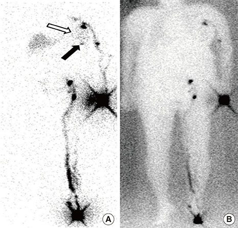
Figure 2 Tc-99m albumin nanocolloid planar lymphoscintigraphy obtained at 3 hours, after a second tracer injection in the left foot. (A) Mild diffuse tracer uptake at the left breast is noted (hollow arrow), compatible with a chyle leak. Another tiny focal tracer uptake (solid arrow) was seen inferiorly. (B) Image with background flood source.
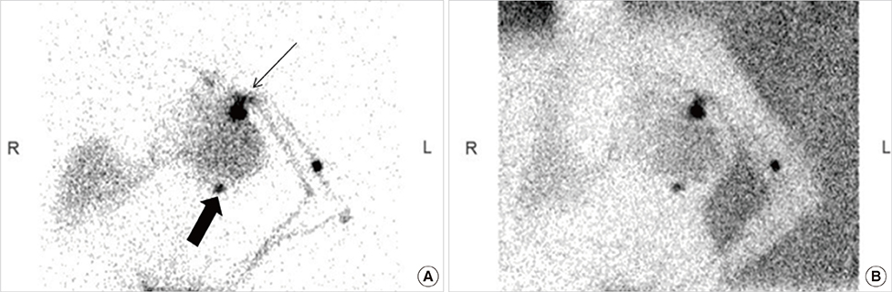
Figure 3 Tc-99m albumin nanocolloid planar lymphoscintigraphy obtained at 4 hours. (A) Delayed images obtained at 4 hours showed increasing tracer intensity at the left axilla (thin arrow) and left breast (thick arrow). (B) Image with background flood source.

Figure 4 Single-photon emission computed tomography/computed tomography (SPECT/CT) scan of the thorax. (A) SPECT/CT reveals focal uptake at the left axilla (arrow). (B) Correlative CT scan localized this uptake to be beyond the left subclavian vein, anterior to the left axillary vein, and inferior to the left biceps tendon (arrow). (C, D) This tracer uptake empties into the chyle collection (arrows). (E, F) Tracer uptake inferior to the chyle collection locates within the collection itself (arrow), as confirmed by correlative CT scan (arrow), likely representing nonuniformity of the chyle concentration.
Reference
-
1. Merrigan BA, Winter DC, O'Sullivan GC. Chylothorax. Br J Surg. 1997; 84:15–20.
Article2. Abdelrazeq AS. Lymphoscintigraphic demonstration of chylous leak after axillary lymph node dissection. Clin Nucl Med. 2005; 30:299–301.
Article3. Pui MH, Yueh TC. Lymphoscintigraphy in chyluria, chyloperitoneum and chylothorax. J Nucl Med. 1998; 39:1292–1296.4. Fitz-Hugh GS, Cowgill R. Chylous fistula. Arch Otolaryngol. 1970; 91:543–547.5. Nakajima E, Iwata H, Iwase T, Murai H, Mizutani M, Miura S, et al. Four cases of chylous fistula after breast cancer resection. Breast Cancer Res Treat. 2004; 83:11–14.
Article6. Singh M, Deo SV, Shukla NK, Pandit A. Chylous fistula after axillary lymph node dissection: incidence, management, and possible cause. Clin Breast Cancer. 2011; 11:320–324.
Article7. Rice DC, Emory RE Jr, McIlrath DC, Meland NB. Chylous fistula: an unusual occurrence after mastectomy with immediate breast reconstruction. Plast Reconstr Surg. 1994; 93:399–401.8. Baek JM, Lee JA, Nam YH, Sung GY, Lee do S, Won JM. Chylous leakage: a rare complication after axillary lymph node dissection in breast cancer and surgical management. J Breast Cancer. 2012; 15:133–134.
Article9. Mariani G, Bruselli L, Kuwert T, Kim EE, Flotats A, Israel O, et al. A review on the clinical uses of SPECT/CT. Eur J Nucl Med Mol Imaging. 2010; 37:1959–1985.
Article10. Shreve PD. Adding structure to function. J Nucl Med. 2000; 41:1380–1382.11. Schillaci O, Simonetti G. Fusion imaging in nuclear medicine: applications of dual-modality systems in oncology. Cancer Biother Radiopharm. 2004; 19:1–10.
Article12. Ohtsuka A, Inoue Y, Asano Y, Woodhams R, Shiomi K. Lymphoscintigraphy using dynamic imaging and SPECT/CT in chylothorax. Open J Med Imaging. 2013; 3:86–89.
Article13. Kotani K, Kawabe J, Higashiyama S, Shiomi S. Lymphoscintigraphy with single-photon emission computed tomography/computed tomography is useful for determining the site of chyle leakage after esophagectomy. Indian J Nucl Med. 2012; 27:208–209.
- Full Text Links
-
- Actions
-
Cited
- CITED
-
- Close
- Share
-
- Similar articles
-
- Sentinel Lymph Node Imaging in Breast Cancer
- Usefulness of Additional SPECT/CT Identifying Lymphatico-renal Shunt in a Patient with Chyluria
- Unusual Contralateral Axillary Lymph Node Metastasis in a Second Primary Breast Cancer Detected by FDG PET/CT and Lymphoscintigraphy
- Use of Mammary Lymphoscintigraphy and Intraoperative Radioguided Gamma Probe in Sentinel Lymph Node Biopsy of Breast Cancer
- Extraordinary Lymph Drainage in Cutaneous Malignant Melanoma and the Value of Hybrid Imaging: A Case Report
