Clin Exp Otorhinolaryngol.
2010 Mar;3(1):24-26.
Classification of External Auditory Canal Cholesteatoma by Computed Tomography
- Affiliations
-
- 1Department of Otorhinolaryngology, Yonsei University College of Medicine, Seoul, Korea.
- 2Soree Ear Clinic, Seoul, Korea. earclinic@hanmail.net
Abstract
OBJECTIVES
We propose here a classification system for external auditory canal cholesteatoma (EACC). We classified the EACC by the computed tomography findings and clinical findings of the patients, and we evaluated the EACC characteristics by the proposed staging system.
METHODS
Stage classification was done according to the results of temporal bone computed tomography and the clinical findings of the patients. Stage I indicates that the EACC lesion is limited to the external auditory canal. Stage II indicates that the EACC lesion invades the tympanic membrane and middle ear. Stage III indicates that the EACC lesion creates a defect of the external auditory canal and it involves the air cells in the mastoid bone. Stage IV indicates that the EACC lesion is beyond the temporal bone. Between 1996 and 2006, 29 patients with EACC and who underwent surgery were prospectively collected. This study was comprised of 16 males and 13 females with a mean age of 22.8+/-15.0 yr. We reviewed the characteristics and results of surgery by our proposed staging system.
RESULTS
A total of 29 patients who underwent operation due to EACC were classified by this system, and the number of stage I, II, III, and IV cases was 14, 3, 10, and 2, respectively. Symptoms such as otorrhea, hearing impairment and otalgia occurred in 12, 17, and 17 cases, respectively. The most common wall invaded by EACC was the inferior wall. The number of cases that had a spontaneous, congenital, post-traumatic, post-inflammatory or tumorous origin was 14, 9, 2, 2, and 1, respectively. Cholesteatoma recurred in 2 patients after surgery. Both cases were stage 1 and both were caused by congenital disease. There were 3 cases with meatal stenosis after surgery, and their primary disease was congenital.
CONCLUSION
This proposed staging is simple and easily applicable for use when deciding the treatment plan for patients with EACC.
Keyword
MeSH Terms
Figure
-
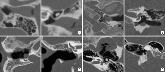
Fig. 1 4 stages of external auditory canal cholesteatoma (EACC) on temporal bone computed tomography. (A) Stage I. The lesion was within the external auditory canal. (B) Stage II. The lesion invaded the middle ear. (C) Stage III. The lesion invaded the mastoid cavity. (D) Stage IV. The lesion extended beyond the temporal bone.
Reference
-
1. Heilbrun ME, Salzman KL, Glastonbury CM, Harnsberger HR, Kennedy RJ, Shelton C. External auditory canal cholesteatoma: clinical and imaging spectrum. AJNR Am J Neuroradiol. 2003; 4. 24(4):751–756. PMID: 12695217.2. Toynbee J. Specimens of molluscum contagiosum developed in the external auditory meatus. Lond Med Gazzette. 1850; 46:811.3. Piepergerdes MC, Kramer BM, Behnke EE. Keratosis obturans and external auditory canal cholesteatoma. Laryngoscope. 1980; 3. 90(3):383–391. PMID: 7359960.
Article4. Naim R, Linthicum F Jr, Shen T, Bran G, Hormann K. Classification of the external auditory canal cholesteatoma. Laryngoscope. 2005; 3. 115(3):455–460. PMID: 15744157.
Article
- Full Text Links
-
- Actions
-
Cited
- CITED
-
- Close
- Share
-
- Similar articles
-
- A Case of Osteoma with Cholesteatoma in the External Auditory Canal
- Clinical Analysis of External Auditory Canal Cholesteatoma
- Distribution of Cytokeratins in Cholesteatomas according to the Induction Period of Cholesteatoma in Gerbil
- Spontaneous migration of a congenital intratympanic membrane cholesteatoma
- First Branchial Cleft Anomaly Presenting External Auditory Canal Atresia and Duplication Anomaly
