Three-dimensional evaluation of the relationship between dental and basal arch forms in normal occlusion
- Affiliations
-
- 1Department of Orthodontics, College of Medicine, The Catholic University of Korea, Korea.
- 2Department of Orthodontics, Graduate School of Clinical Dental Science, The Catholic University of Korea, Korea.
- 3Department of Orthodontics, St. Vincent Hospital, The Catholic University of Korea, Korea.
- 4Department of Orthodontics, School of Dentistry, Seoul National University, Korea.
- 5Department of Orthodontics, Seoul St. Mary's Hospital, The Catholic University of Korea, Korea. kook2002@catholic.ac.kr
- KMID: 2274263
- DOI: http://doi.org/10.4041/kjod.2011.41.4.288
Abstract
OBJECTIVE
The purposes of this study were to evaluate the relationship between the dental and basal arch forms; to analyze their differences in the tapered, ovoid, and square arch forms in normal occlusion by using three-dimensional (3D) virtual models; and to test the hypothesis that the overjet and maxillomandibular basal arch width difference have a significantly positive correlation.
METHODS
Seventy-seven normal occlusion plaster casts were examined by 3D scanning. Facial axis (FA) and WALA points were digitized using the Rapidform 2006 software. The dimensions of the dental and basal arches and the overjet were measured. The samples were classified into 3 groups according to arch forms: tapered (n = 20), ovoid (n = 20), and square (n = 37). Analysis of variance (ANOVA) was used to compare the dental and basal arch dimensions. The Pearson correlation coefficients between the intercanine as well as the intermolar widths at the FA and WALA points were calculated.
RESULTS
With regard to the basal arch dimensions, the tapered arch form showed a larger mandibular intermolar depth than the ovoid. Strong correlations were noted between the basal and dental intermolar widths in both the upper and lower arches (r = 0.83 and 0.85, respectively). Moderate correlation was found between the upper and lower intercanine widths (r = 0.65 and 0.48, respectively).
CONCLUSIONS
The 3 dental arch form groups differed only in some dimensions of the skeletal arch. Moderate correlations were found between the basal and dental intercanine widths. These findings suggest that the basal arch may not be a principle factor in determining the dental arch form.
Keyword
Figure
-

Fig. 1 FA (green) and WALA (purple) points defined on a virtual model and seen in the A, Upper front, B, lower front, C, upper buccal, and D, lower buccal views. FA, Facial axis; WALA, Will Andrews and Larry Andrews.
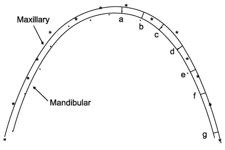
Fig. 2 Overjet measured as the shortest distance from each FA (facial axis) point on the mandibular arch to that on the maxillary arch. a, Central incisor; b, lateral incisor; c, canine; d, first premolar; e, second premolar; f, first molar; g, second molar.

Fig. 3 Average relative distances between corresponding WALA and FA points in the upper and lower arches. WALA, Will Andrews and Larry Andrews; FA, facial axis.
Cited by 1 articles
-
Comparison between dental and basal arch forms in normal occlusion and Class III malocclusions utilizing cone-beam computed tomography
Kyung Eun Suk, Jae Hyun Park, Mohamed Bayome, Young-Ok Nam, Glenn T. Sameshima, Yoon-Ah Kook
Korean J Orthod. 2013;43(1):15-22. doi: 10.4041/kjod.2013.43.1.15.
Reference
-
1. Betts NJ, Vanarsdall RL, Barber HD, Higgins-Barber K, Fonseca RJ. Diagnosis and treatment of transverse maxillary deficiency. Int J Adult Orthodon Orthognath Surg. 1995. 10:75–96.2. Lundström AF. Malocclusion of the teeth regarded as a problem in connection with the apical base. Int J Orthod Oral Surg Radiogr. 1925. 9:591–602. 724–731. 793–812. 933–940. 1022–1042. 1109–1133.
Article3. Proffit WR, Fields HW, Sarver DM. Proffit WR, Fields HW, Sarver DM, editors. Orthodontic treatment planning: limitations, controversies, and special problems. Contemporary orthodontics. 2000. St Louis: Mosby;276–279.4. Strang RHW. The fallacy of denture expansion as a treatment procedure. Angle Orthod. 1949. 19:12–22.5. Handelman CS. The anterior alveolus: its importance in limiting orthodontic treatment and its influence on the occurrence of iatrogenic sequelae. Angle Orthod. 1996. 66:95–109.6. Johnson KC. Cases six years postretention. Angle Orthod. 1977. 47:210–221.7. Little RM, Wallen TR, Riedel RA. Stability and relapse of mandibular anterior alignment-first premolar extraction cases treated by traditional edgewise orthodontics. Am J Orthod. 1981. 80:349–365.
Article8. Shapiro PA. Mandibular dental arch form and dimension. Treatment and postretention changes. Am J Orthod. 1974. 66:58–70.9. Carmen M, Marcella P, Giuseppe C, Roberto A. Periodontal evaluation in patients undergoing maxillary expansion. J Craniofac Surg. 2000. 11:491–494.
Article10. Vanarsdall RL. Graber TM, Swain BF, editors. Periodontal/orthodontic interrelationships. Orthodontics, current principles and techniques. 1994. St Louis: Mosby;712–749.11. Vanarsdall RL Jr. Transverse dimension and long-term stability. Semin Orthod. 1999. 5:171–180.
Article12. Vanarsdall RL, White RP Jr. Three-dimensional analysis for skeletal problems. Int J Adult Orthodon Orthognath Surg. 1994. 9:159.13. Andrews LF, Andrews WA. The six elements of orofacial harmony. Andrews J. 2000. 1:13–22.14. Ball RL, Miner RM, Will LA, Arai K. Comparison of dental and apical base arch forms in Class II Division1 and Class I malocclusions. Am J Orthod Dentofacial Orthop. 2010. 138:41–50.
Article15. Gupta D, Miner RM, Arai K, Will LA. Comparison of the mandibular dental and basal arch forms in adults and children with Class I and Class II malocclusions. Am J Orthod Dentofacial Orthop. 2010. 138:10.e1–10.e8.
Article16. Ronay V, Miner RM, Will LA, Arai K. Mandibular arch form: the relationship between dental and basal anatomy. Am J Orthod Dentofacial Orthop. 2008. 134:430–438.
Article17. Kim SC. A study on the configurations of Korean normal dental arches for preformed arch wire. Korean J Orthod. 1984. 14:93–101.18. Kook YA, Nojima K, Moon HB, McLaughlin RP, Sinclair PM. Comparison of arch forms between Korean and North American white populations. Am J Orthod Dentofacial Orthop. 2004. 126:680–686.
Article19. Merz ML, Isaacson RJ, Germane N, Rubenstein LK. Tooth diameters and arch perimeters in a black and a white population. Am J Orthod Dentofacial Orthop. 1991. 100:53–58.
Article20. Nojima K, McLaughlin RP, Isshiki Y, Sinclair PM. A comparative study of Caucasian and Japanese mandibular clinical arch forms. Angle Orthod. 2001. 71:195–200.21. Nummikoski P, Prihoda T, Langlais RP, McDavid WD, Welander U, Tronje G. Dental and mandibular arch widths in three ethnic groups in Texas: a radiographic study. Oral Surg Oral Med Oral Pathol. 1988. 65:609–617.
Article22. Yun YK, Kook YA, Kim SH, Mo SS, Cha KS, Kim JG, et al. Mandibular clinical arch forms in Koreans with normal occlusions. Korean J Orthod. 2004. 34:481–487.23. Bayome M, Sameshima GT, Kim Y, Nojima K, Baek SH, Kook YA. Comparison of arch form between Egyptian and North American white populations. Am J Orthod Dentofacial Orthop. 2011. 139:e245–e252.24. Cordato MA. A simple mathematical study of anterior dental relations: Part I. Aust Orthod J. 1995. 13:249–252.25. Cordato MA. A mathematical study of anterior dental relations: Part II, Incisor and canine overjet. Aust Orthod J. 1996. 14:143–149.26. Cordato MA. A simple mathematical study of anterior dental relations: Part III: incisor and canine overbite. Aust Orthod J. 1998. 15:75–84.27. Ferrario VF, Sforza C, Miani A Jr, Tartaglia G. Maxillary versus mandibular arch form differences in human permanent dentition assessed by Euclidean-distance matrix analysis. Arch Oral Biol. 1994. 39:135–139.
Article28. Ferrario VF, Sforza C, Miani A Jr, Tartaglia G. Mathematical definition of the shape of dental arches in human permanent healthy dentitions. Eur J Orthod. 1994. 16:287–294.
Article29. Lee YC, Park YC. A study on the dental arch by occlusogram in normal occlusion. Korean J Orthod. 1987. 17:279–287.30. Kook YA, Bayome M, Park SB, Cha BK, Lee YW, Beck SH. Overjet at the anterior and posterior segments: three-dimensional analysis of arch coordination. Angle Orthod. 2009. 79:495–501.
Article31. Triviño T, Siqueira DF, Scanavini MA. A new concept of mandibular dental arch forms with normal occlusion. Am J Orthod Dentofacial Orthop. 2008. 133:10.e15–10.e22.
Article32. Kim BI, Bayome M, Kim Y, Baek SH, Han SH, Kim SH, et al. Comparison of overjet among 3 arch types in normal occlusion. Am J Orthod Dentofacial Orthop. 2011. 139:e253–e260.
Article33. Andrews LF. The six keys to normal occlusion. Am J Orthod. 1972. 62:296–309.
Article34. Felton JM, Sinclair PM, Jones DL, Alexander RG. A computerized analysis of the shape and stability of mandibular arch form. Am J Orthod Dentofacial Orthop. 1987. 92:478–483.
Article35. Pepe SH. Polynomial and catenary curve fits to human dental arches. J Dent Res. 1975. 54:1124–1132.
Article36. Fujita K, Takada K, QianRong G, Shibata T. Patterning of human dental arch wire blanks using a vector quantization algorithm. Angle Orthod. 2002. 72:285–294.37. Sergl HG, Kerr WJ, McColl JH. A method of measuring the apical base. Eur J Orthod. 1996. 18:479–483.
Article38. Tweed CH. A philosophy of orthodontic treatment. Am J Orthod Oral Surg. 1945. 31:74–103.
Article39. Hesby RM, Marshall SD, Dawson DV, Southard KA, Casko JS, Franciscus RG, et al. Transverse skeletal and dentoalveolar changes during growth. Am J Orthod Dentofacial Orthop. 2006. 130:721–731.
Article40. Quimby ML, Vig KW, Rashid RG, Firestone AR. The accuracy and reliability of measurements made on computer-based digital models. Angle Orthod. 2004. 74:298–303.41. Zilberman O, Huggare JV, Parikakis KA. Evaluation of the validity of tooth size and arch widthmeasurements using conventional and three-dimensional virtual orthodotic models. Angle Orthod. 2003. 73:301–306.42. Costalos PA, Sarraf K, Cangialosi TJ, Efstratiadis S. Evaluation of the accuracy of digital model analysis for the American Board of Orthodontics objective grading system for dental casts. Am J Orthod Dentofacial Orthop. 2005. 128:624–629.
Article43. Sangcharearn Y, Ho C. Effect of incisor angulation on overjet and overbite in class II camouflage treatment. A typodont study. Angle Orthod. 2007. 77:1011–1018.
Article44. Lowe DW, Forbes DB, Hicken J, Skarin D. The effect of labiolingual tooth dimension on proper overbite and overjet. Northwest Dent Res. 1997. 7:31–43.
- Full Text Links
-
- Actions
-
Cited
- CITED
-
- Close
- Share
-
- Similar articles
-
- Three dimensional structural analysis between dental arch and basal bone in normal occlusion
- Comparative analysis of the relationship between basal bone and teeth in normal occlusion and Angle's Class I malocclusion
- Comparison between dental and basal arch forms in normal occlusion and Class III malocclusions utilizing cone-beam computed tomography
- A study on the dental arch by occlusogram in normal occlusion
- The new approach to maxillary and mandibular anterior dental arch forms: The prediction to maxillary and mandibular anterior occlusal relationship by computer program
