Korean J Orthod.
2014 Jan;44(1):44-49. 10.4041/kjod.2014.44.1.44.
Effects of ultrasonic instrumentation with different scaler-tip angulations on the shear bond strength and bond failure mode of metallic orthodontic brackets
- Affiliations
-
- 1Department of Orthodontics, University of Bologna, Bologna, Italy. giulio.alessandri@unibo.it
- 2Department of Periodontology, University of Bologna, Bologna, Italy.
- KMID: 2273270
- DOI: http://doi.org/10.4041/kjod.2014.44.1.44
Abstract
OBJECTIVE
To evaluate the effects of ultrasonic instrumentation with different scaler-tip angulations on the shear bond strength (SBS) and bond failure mode of metallic orthodontic brackets.
METHODS
Adhesive pre-coated metallic brackets were bonded to 72 extracted human premolars embedded in autopolymerizing acrylic resin. The teeth were randomly divided into 3 groups (n = 24 each) to undergo no treatment (control group) or ultrasonic instrumentation with a scaler-tip angulation of 45degrees (45degrees-angulation group) or 0degrees (0degrees-angulation group). SBS was tested in a universal testing machine, and adhesive remnant index (ARI) scores were recorded. The Kruskal-Wallis test and Mann-Whitney U-test were used for statistical analysis.
RESULTS
The control group had a significantly higher mean SBS value than the treated groups, which showed no significant differences in their mean SBS values. The ARI scores were not significantly different among the groups.
CONCLUSIONS
Ultrasonic instrumentation around the bracket base reduces the SBS of metallic orthodontic brackets, emphasizing the need for caution during professional oral hygiene procedures in orthodontic patients. The scaler-tip angulation does not influence the SBS reduction and bond failure mode of such brackets.
Keyword
Figure
-
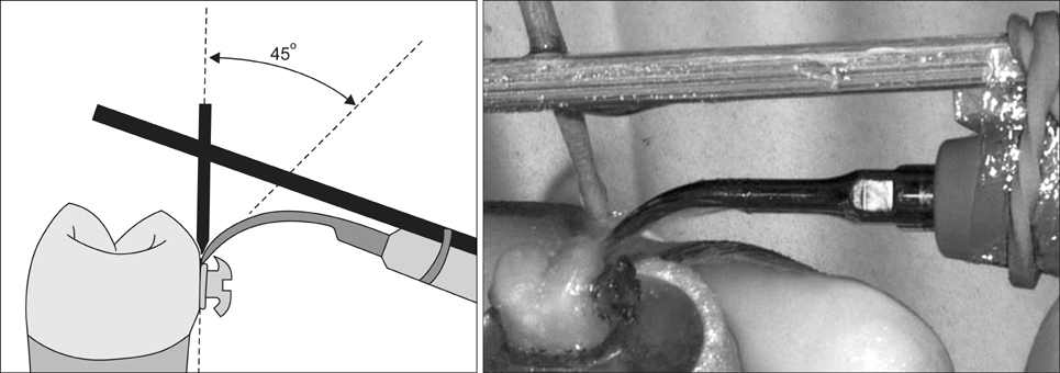
Figure 1 Ultrasonic instrumentation with the scaler-tip angulation of 45°.
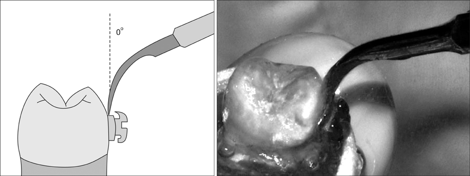
Figure 2 Ultrasonic instrumentation with the scaler-tip angulation of 0°.
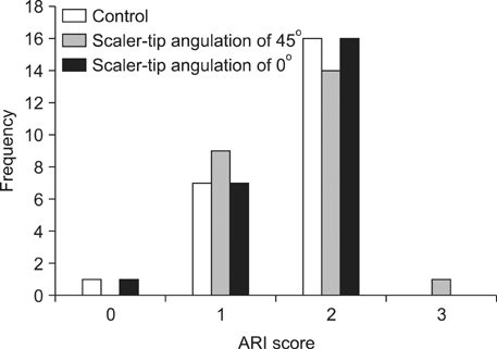
Figure 3 Distribution of the adhesive remnant index (ARI) scores.
Reference
-
1. Ogaard B, Rølla G, Arends J. Orthodontic appliances and enamel demineralization. Part 1. Lesion development. Am J Orthod Dentofacial Orthop. 1988; 94:68–73.2. Zanarini M, Pazzi E, Bonetti S, Ruggeri O, Alessandri Bonetti G, Prati C. In vitro evaluation of the effects of a fluoride-releasing composite on enamel demineralization around brackets. Prog Orthod. 2012; 13:10–16.
Article3. Chapman JA, Roberts WE, Eckert GJ, Kula KS, González-Cabezas C. Risk factors for incidence and severity of white spot lesions during treatment with fixed orthodontic appliances. Am J Orthod Dentofacial Orthop. 2010; 138:188–194.
Article4. Atack NE, Sandy JR, Addy M. Periodontal and microbiological changes associated with the placement of orthodontic appliances. A review. J Periodontol. 1996; 67:78–85.
Article5. Petti S, Barbato E, Simonetti D'Arca A. Effect of orthodontic therapy with fixed and removable appliances on oral microbiota: a six-month longitudinal study. New Microbiol. 1997; 20:55–62.6. Ristic M, Vlahovic Svabic M, Sasic M, Zelic O. Clinical and microbiological effects of fixed orthodontic appliances on periodontal tissues in adolescents. Orthod Craniofac Res. 2007; 10:187–195.
Article7. Park SY, Cha JY, Kim KN, Hwang CJ. The effect of casein phosphopeptide amorphous calcium phosphate on the in vitro shear bond strength of orthodontic brackets. Korean J Orthod. 2013; 43:23–28.
Article8. Checchi L, Nobili A. The importance of hygiene control in orthodontics. Prev Assist Dent. 1988; 14:35–37.9. Gwinnett AJ, Ceen RF. Plaque distribution on bonded brackets: a scanning microscope study. Am J Orthod. 1979; 75:667–677.
Article10. Sukontapatipark W, el-Agroudi MA, Selliseth NJ, Thunold K, Selvig KA. Bacterial colonization associated with fixed orthodontic appliances. A scanning electron microscopy study. Eur J Orthod. 2001; 23:475–484.
Article11. Olin PS. Effect of prolonged ultrasonic instrumentation on the retention of cemented cast crowns. J Prosthet Dent. 1990; 64:563–565.
Article12. Krell KV, Courey JM, Bishara SE. Orthodontic bracket removal using conventional and ultrasonic debonding techniques, enamel loss, and time requirements. Am J Orthod Dentofacial Orthop. 1993; 103:258–266.
Article13. Boyer DB, Engelhardt G, Bishara SE. Debonding orthodontic ceramic brackets by ultrasonic instrumentation. Am J Orthod Dentofacial Orthop. 1995; 108:262–266.
Article14. Bishara SE, Trulove TS. Comparisons of different debonding techniques for ceramic brackets: an in vitro study. Part I. Background and methods. Am J Orthod Dentofacial Orthop. 1990; 98:145–153.
Article15. Bishara SE, Trulove TS. Comparisons of different debonding techniques for ceramic brackets: an in vitro study. Part II. Findings and clinical implications. Am J Orthod Dentofacial Orthop. 1990; 98:263–273.
Article16. Walmsley AD, Jones PA, Hullah W, Harrington E. Ultrasonic debonding of composite-retained restorations. Br Dent J. 1989; 166:290–294.
Article17. Giulio AB, Matteo Z, Serena IP, Silvia M, Luigi C. In vitro evaluation of casein phosphopeptide-amorphous calcium phosphate (CPP-ACP) effect on stripped enamel surfaces. A SEM investigation. J Dent. 2009; 37:228–232.
Article18. International Organization for Standardization. ISO/TS 11405:2003. Dental materials - Testing of adhesion to tooth structure. ISO;2003. cited Apr 23, 2013. Available from: http://www.iso.org/iso/home/store/catalogue_tc/catalogue_detail.htm?csnumber=31486.19. Alessandri Bonetti G, Zanarini M, Incerti Parenti S, Lattuca M, Marchionni S, Gatto MR. Evaluation of enamel surfaces after bracket debonding: an in-vivo study with scanning electron microscopy. Am J Orthod Dentofacial Orthop. 2011; 140:696–702.
Article20. Vicente A, Bravo LA, Romero M, Ortiz AJ, Canteras M. Adhesion promoters: effects on the bond strength of brackets. Am J Dent. 2005; 18:323–326.21. Montasser MA, Drummond JL. Reliability of the adhesive remnant index score system with different magnifications. Angle Orthod. 2009; 79:773–776.
Article22. Artun J, Bergland S. Clinical trials with crystal growth conditioning as an alternative to acid-etch enamel pretreatment. Am J Orthod. 1984; 85:333–340.
Article23. Pont HB, Özcan M, Bagis B, Ren Y. Loss of surface enamel after bracket debonding: an in-vivo and ex-vivo evaluation. Am J Orthod Dentofacial Orthop. 2010; 138:387.e1–387.e9. discussion 387-9.
Article24. Müller P, Guggenheim B, Attin T, Marlinghaus E, Schmidlin PR. Potential of shock waves to remove calculus and biofilm. Clin Oral Investig. 2011; 15:959–965.
Article25. Chapple IL, Walmsley AD, Saxby MS, Moscrop H. Effect of instrument power setting during ultrasonic scaling upon treatment outcome. J Periodontol. 1995; 66:756–760.
Article26. Reynolds IR. A review of direct orthodontic bonding. Br J Orthod. 1975; 2:171–180.
Article27. Lombardo L, Kaplan A, Lapenta R, Bratti E, Pera C, Scuzzo G, et al. A comparative study of lingual bracket bond strength. Orthodontics (Chic.). 2011; 12:178–187.28. Arabaci T, Ciçek Y, Canakçi CF. Sonic and ultrasonic scalers in periodontal treatment: a review. Int J Dent Hyg. 2007; 5:2–12.
Article29. Flemmig TF, Petersilka GJ, Mehl A, Hickel R, Klaiber B. The effect of working parameters on root substance removal using a piezoelectric ultrasonic scaler in vitro. J Clin Periodontol. 1998; 25:158–163.
Article
- Full Text Links
-
- Actions
-
Cited
- CITED
-
- Close
- Share
-
- Similar articles
-
- The shear bond strength of two adhesives bonded to composite resin and glass ionomer cement restorations
- Effect of applying adhesive after enamel etching on the shear bond strength of orthodontic brackets using light curing resin cements
- A change of shear bond strength of orthodontic resin adhesives under water immersion
- The shear bond strength and adhesive failure pattern in bracket bonding with different light-curing methods
- A study on the shear bond strengths of orthodontic brackets according to surface treatments and sizes of amalgam restorations.
