Ann Rehabil Med.
2011 Oct;35(5):719-724. 10.5535/arm.2011.35.5.719.
The Shoulder Gradient in Patients with Unilateral Shoulder Impingement Syndrome
- Affiliations
-
- 1Department of Physical Medicine and Rehabilitation, Kyung Hee University College of Medicine, Seoul 130-702, Korea. daegyuhwang@gmail.com
- KMID: 2267235
- DOI: http://doi.org/10.5535/arm.2011.35.5.719
Abstract
OBJECTIVE
To investigate the relationship between the shoulder gradient and acromiohumeral interval of both shoulders in patients with unilateral shoulder impingement syndrome. METHOD: Using the angulometer, we measured the shoulder gradient in patients with unilateral shoulder impingement syndrome in a standing position. Using the radiography, we measured the acromiohumeral interval and the angle between a vertical line and a line connecting a superior angle with an inferior angle of the scapula.
RESULTS
In patients with unilateral shoulder impingement syndrome, the frequency of shoulder impingement syndrome was 76.2% (16 of 21) on the side of the relatively lower shoulder. The mean acromiohumeral interval on the side of the lower shoulder was 10.03+/-1.28 mm, compared with 10.46+/-1.50 mm for the higher shoulder. The angle between a vertical line and a line connecting a superior angle with an inferior angle of the scapular of the side of the lower shoulder was -0.31+/-3.73 degrees, compared with 3.85+/-4.42 degrees for the higher shoulder.
CONCLUSION
The frequency of shoulder impingement syndrome was significantly higher on the side of the relatively lower shoulder, and there is no significant difference in the acromiohumeral interval between the side of the lower shoulder and that of the higher shoulder. In patients with unilateral shoulder impingement syndrome, the scapular on the side of lower shoulder was more rotated downward than on the side of the higher shoulder.
MeSH Terms
Figure
-
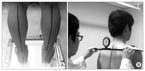
Fig. 1 Patient stood on the scaled podoscopesith ankles in neutral positionand knees are full extended (A). Shoulder gradoent was measured byangulmeter with each side of blade on patient's most lateral part of acromion (B).
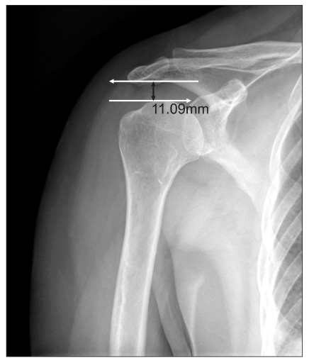
Fig. 2 This AP radiograph shows the acromiohumeral interval, which was measured as the shortest distance from the inferior surface of the acromion to the superior aspect of the humerus.
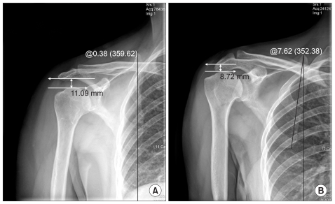
Fig. 3 This AP radiograph shows upward and downward rotation of scapula, which was measured as the angle between the vertical line and the line connecting a superior angle with an inferior angle of the scapula. Upward rotation of scapula (A), downward rotation of scapula (B).
Reference
-
1. Ogata S, Uhthoff HK. Acromial enthesopathy and rotator cuff tear. A radiologic and histologic postmortem investigation of the coracoacromial arch. Clin Orthop Relat Res. 1990; 254:39–48. PMID: 2323148.2. Neer CS. Impingment lesions. Clin Orthop Relat Res. 1983; 173:70–77. PMID: 6825348.3. Schamberger W. Schamberger W, editor. The malalignment syndrome. The malalignment syndrome. 2002. 1st ed. New York: Churchill Livingstone;p. 106–110.4. Shibuta H, Tamai K, Tabuchi K. Magnetic resonance imaging of the shoulder in abduction. Clin Orthop Relat Res. 1998; 348:107–113. PMID: 9553541.
Article5. Tirman PF, Steinbach LS, Belzer JP, Bost FW. A practical approach to imaging of the shoulder with emphasis on MR imaging. Orthop Clin North Am. 1997; 28:483–515. PMID: 9257962.
Article6. Allmann KH, Uhl M, Gufler H, Biebow N, Hauer MP, Kotter E, Reichelt A, Langer M. Cine-MR imaging of the shoulder. Acta Radiol. 1997; 38:1043–1046. PMID: 9394666.
Article7. Hijioka A, Suzuki K, Nakamura T, Hojo T. Degenerative change and rotator cuff tears. An anatomical study in 160 shoulders of 80 cadavers. Arch Orthop Trauma Surg. 1993; 112:61–64. PMID: 8457412.8. Schneeberger AG, Nyffeler RW, Gerber C. Structural changes of the rotator cuff caused by experimental subacromial impingement in the rat. J Shoulder Elbow Surg. 1998; 7:375–380. PMID: 9752647.
Article9. Rathbun JB, Macnab I. The microvascular pattern of the rotator cuff. J Bone Joint Surg Br. 1970; 52:540–553. PMID: 5455089.
Article10. Ozaki J, Fuzimoto S, Nakagawa Y, Masuhara K, Tamai S. Tears of the rotator cuff of the shoulder associated with pathological changes in the acromion. A study in cadevara. J Bone Joint Surg Am. 1988; 70:1224–1230. PMID: 3417708.11. Petersson CJ, Redlund-Johnell I. The subacromial space in normal radiographs. Acta Orthop Scand. 1984; 55:57–58. PMID: 6702430.12. Norwood LA, Barrack R, Jacobson KE. Clinical presentation of complete tears of the rotator cuff. J Bone Joint Surg Am. 1989; 71:499–505. PMID: 2703509.
Article13. Park TS, Rha JD, Kim YH, Yang KH, Choi SM. A study of the measurement of the distance of the subacromial space in normal shoulder. Korean J Sports Med. 1994; 12:103–108.14. Brossmann J, Preidler KW, Pedowitz RA, White LM, Trudell D, Resnick D. Shoulder impingement syndrome: influence of shoulder position on rotator cuff impingement-an anatomic study. AJR Am J Roentgenol. 1996; 167:1511–1515. PMID: 8956588.
Article15. Solem-Bertoft E, Thuomas KA, Westerberg CE. The influence of scapular retraction and protraction on the width of the subacromial space. An MRI study. Clin Orthop Relat Res. 1993; 296:99–103. PMID: 8222458.16. Nove-Josserand L, Edwards TB, O'Connor DP, Walch G. The acromiohumeral and coracohumeral intervals are abnormal in rotator cuff tears with muscular fatty degeneration. Clin Orthop Relat Res. 2005; 433:90–96. PMID: 15805942.17. Shiri R, Varonen H, Heliövaara M, Viikari-Juntura E. Hand dominance in upper extremity musculoskeletal disorders. J Rheumatol. 2007; 34:1076–1082. PMID: 17343320.
- Full Text Links
-
- Actions
-
Cited
- CITED
-
- Close
- Share
-
- Similar articles
-
- Evaluation of Shoulder Pain in Outpatient Clinic
- Microinstability of the Shoulder
- Changes of Ranges of Motion According to Ages and Manifestation Frequency of Impingement Sign in Shoulder Impingement Syndrome
- Shoulder Impingement Syndrome Combined with Myofascial Pain Syndrome
- Ossification of the Coracoacromial Ligament in Subacromial Impingement Syndrome: A Case Report
