Acquired Tracheoesophageal Fistula through Esophageal Diverticulum in Patient Who Had a Prolonged Tracheostomy Tube: A Case Report
- Affiliations
-
- 1Department of Physical Medicine and Rehabilitation, Kwandong University College of Medicine, Goyang 412-270, Korea. ykkim@kwandong.ac.kr
- KMID: 2266866
- DOI: http://doi.org/10.5535/arm.2011.35.3.436
Abstract
- Acquired tracheoesophageal fistula through esophageal diverticulum is infrequent. We report tracheoesophageal fistula through esophageal diverticulum in a 55-year-old male who had a prolonged tracheostomy tube during 6 months, and a NG tube during 18 months. He suffered from recurrent pneumonia. He complained of a cough associated with eating, and production of sputum mixed with food. To help evaluate the aspiration to the lung and the cause of aspiration, he was tested using gastrointestinal scintigraphy (gastric emptying study), a chest CT scan (pre & post contrast), and esophagoduodenoscopy. The chest CT scan revealed an acquired tracheoesophageal fistula through esophageal diverticulum, and esophagoduodenoscopy revealed a 3 mm sized fistula that was located -33 cm from the upper incisor. We treated the tracheoesophageal fistula by clipping under esophagoduodenoscopy. The symptoms of fever, cough, and aspiration were no long observed after the clipping was completed.
MeSH Terms
Figure
-

Fig. 1 Tc99m-DTPA was swallowed and serial images were obtained. Gastrointestinal scintigraphy revealed faint tracer activity in right side of mid esophagus (Arrow: faint tracer activity). A tracheoesophageal fistula was suspected in this study.
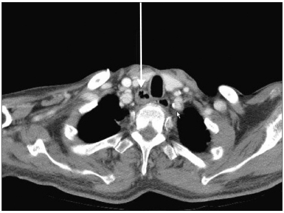
Fig. 2 Transverse chest CT scan revealed the lower neck area mid-esophagus. About 1.8 cm size cavitary lesion was noted in right lower neck at the level of thoracic inlet (arrow). The esophagus was communicated with trachea.

Fig. 3 Esophagoduodenoscopic images: About the 3 mm-sized tracheoesophageal fistula was seen. And also the bubble from the trachea was observed. The fistula was located in the esophagus about 33 cm from the upper incisor (A). Post endoscopic clipping. Endoscopic clipping was done by using 4 clips (B).
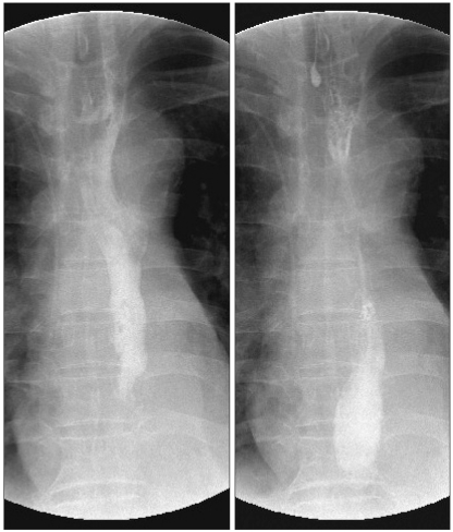
Fig. 4 Endoscopic clipping was done. After 3 days, upper GI series by using gastrograffin revealed that the leakage from mid esophagus was not observed.
Reference
-
1. Kim KH, Lee JS, Yang HW, Kim JH, Kim GH, Lee MS, Kim WJ. A case of tracheoesophageal fistula through esophageal diverticulm combined with aspiration pneumonia. Korean J Gastroenterol. 1997; 30:677–683.2. Hilgenberg AD, Grillo HC. Acquired nonmalignant tracheoesophageal fistula. J Thorac Cardiovasc Surg. 1983; 85:492–498. PMID: 6834870.
Article3. Jung YH, Baek CW, Park JW, Woo YC, Koo GH. Acquired-tracheoesophageal fistula observed after ventilator care. Korean J Anesthesiol. 2004; 46:122–126.5. Kim SC, Ha KW, Wang JH, Kim SJ, Kim WH, Jeong SH, Lee WS, Han SD, Chon GR. Two cases of postintubation tracheoesophageal fistula in patients with a history of tracheostomy. Korean J Crit Care Med. 2009; 24:87–91.6. Paik HC, Kim DH, Cho HM, Lee DY. Clinical analysis of acquired tracheoesophageal fistula. Korean J Bronchoesophagol. 2002; 8:61–65.7. Bertelson S. Congenital oesophago-tracheal fistulas in the adult. Scand J thorac Cardiovasc Surg. 1970; 4:80–82. PMID: 5424780.8. Yeh CM, Chou CM. Early repair of acquired tracheoesophageal fistula. Asian Cardiovasc Thorac Ann. 2008; 16:318–320. PMID: 18670027.
Article9. Jeon SH, Park SW, Jung SW, Lee HR. Postintubation tracheo-esophageal fistula. Korean J Thorac Cardiovasc Surg. 1996; 29:235–238.
- Full Text Links
-
- Actions
-
Cited
- CITED
-
- Close
- Share
-
- Similar articles
-
- Tracheoesophageal Fistula Following Prolonged Tracheostomy Cannulation
- Tracheoesophageal Fistula with Subglottic Stenosis in Tracheostomy Patient: Report of 1 Case
- Repair of Tracheoesophageal Fistula under Laryngeal Microsurgery Approach: Case Report and Literature Review
- Tracheoesophageal Fistula with Tracheal Dilatation in a Patient with a Tracheostomy Using a Home Mechanical Ventilator
- A Case of the Esophageal Atresia with Distal Tracheoesophageal Fistula Associated with Duodenal Obstrction
