Magnetic Resonance Imaging as a Determinant for Surgical Release of Congenital Muscular Torticollis: Correlation with the Histopathologic Findings
- Affiliations
-
- 1The Center for Torticollis, Department of Physical Medicine and Rehabilitation, Ajou University School of Medicine, Suwon 443-721, Korea. syyim@ajou.ac.kr
- 2Department of Pathology, Ajou University School of Medicine, Suwon 443-721, Korea.
- 3Department of Plastic and Reconstructive Surgery, Ajou University School of Medicine, Suwon 443-721, Korea.
- 4Department of Radiology, Ajou University School of Medicine, Suwon 443-721, Korea.
- KMID: 2266736
- DOI: http://doi.org/10.5535/arm.2012.36.3.320
Abstract
OBJECTIVE
(1) To present the magnetic resonance imaging (MRI) findings of congenital muscular torticollis (CMT) of subjects who underwent surgical release and subjects who showed a good prognosis with stretching exercises and (2) to correlate the MRI findings with the histopathologic findings of CMT for subjects who underwent surgical release in order to examine the hypothesis that the MRI findings of CMT can be used as a determinant to perform surgical release of CMT. METHOD: The neck MRI findings of 33 subjects who underwent surgical release for CMT were compared with those of 18 subjects who were successfully managed only with conservative management. The MRI findings were correlated with the histopathologic sections of the CMT mass.
RESULTS
All 33 subjects (100%) who underwent surgical release showed one or more low signal intensities within the involved sternocleidomastoid muscle (SCM) on the T1- and T2-weighted images of neck MRI. The eighteen non-surgical candidates showed only enlargement of the SCM without low signal intensity within the SCM. The histopathologic findings showed interstitial fibrosis and/or the presence of aberrant tendon-like excessive dense connective tissue that was either well-arranged or disorganized.
CONCLUSION
The histopathologic findings and MRI findings showed good correlation in terms of the amount of fibrosis and aberrant dense connective tissue within the SCM. If multiple or large low signal intensities within the SCM are noted, we think that surgical release should be considered.
MeSH Terms
Figure
-
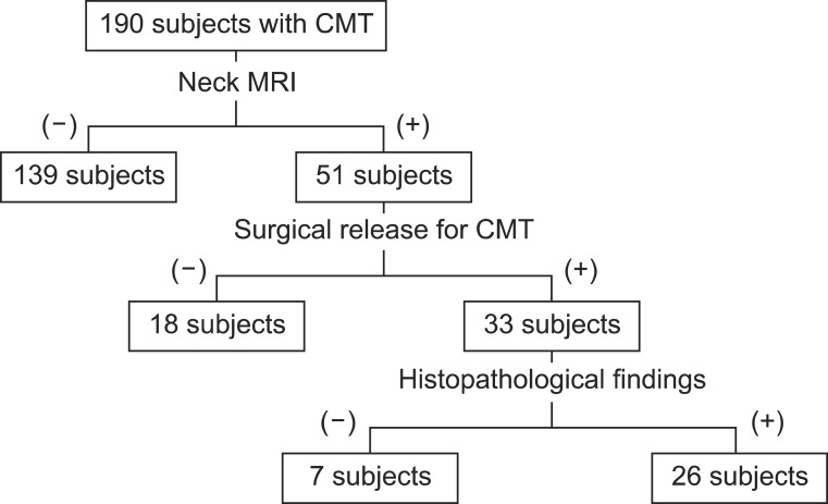
Fig. 1 The algorithm for enrollment of subjects from January 2009 to October 2009.
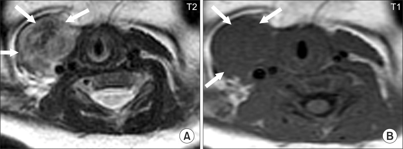
Fig. 2 The neck MRI findings of a one month old girl with right congenital muscular torticollis show high signal intensity on the (A) T2-weighted image compared to the (B) T1-weighted image (arrows).
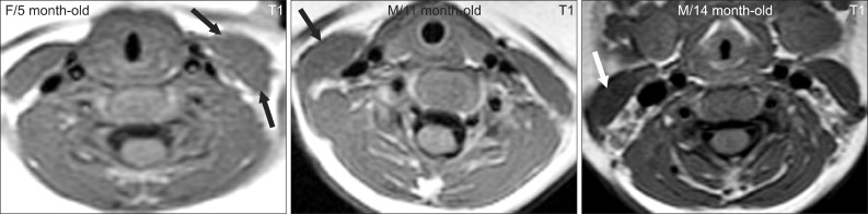
Fig. 3 Typical neck MRI findings of subjects who show a good response to stretching exercises. Thickened sternocleidomastoid muscles do not have low signal intensities (arrows).
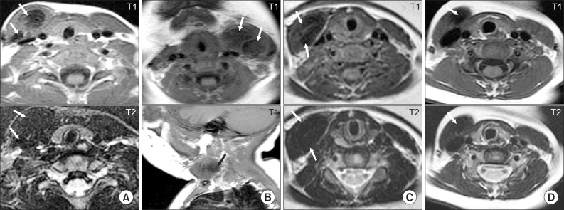
Fig. 4 Typical neck MRI findings of subjects who underwent surgical release for congenital muscular torticollis (CMT). (A) Eleven month old boy with right CMT showing low signal intensities on the T1- and T2-weighted axial images of both the sternal head and clavicular head (arrows) of the right sternocleidomastoid muscle (SCM). (B) Four month old girl with left CMT showing low signal intensities on the T1-weighted axial and sagittal images of the left SCM (arrows). (C) Six month old girl with right CMT showing low signal intensities on both the T1- and T2-weighted axial images of the right SCM (arrows). (D) Eighteen month old girl with right CMT showing low signal intensity on both the T1- and T2-weighted axial images of the right SCM (arrows).
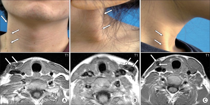
Fig. 5 Pictures showing subjects with atrophy or same thickness of the shortened sternocleidomastoid muscle (SCM) with low signal intensities on the T1-weighted axial images of the SCM. (A) Twenty-two year old man with right congenital muscular torticollis (CMT) showing a cord-like right SCM (arrows). Neck MRI showed low signal intensities (arrows) and did not show significant difference of thickness between the right and left SCM. (B) Five year old girl with left CMT showing a cord-like left SCM (arrows) and low signal intensity (arrows) on left SCM. (C) Twenty year old woman with right CMT showing atrophied cord-like right SCM (arrows) with low signal intensity (arrows) on right SCM.
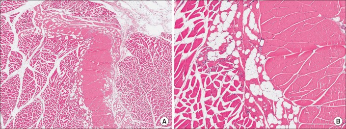
Fig. 6 The histologic findings of the normal sternocleidomastoid muscle without congenital muscular torticollis (H&E, (A) ×100; (B) ×200).
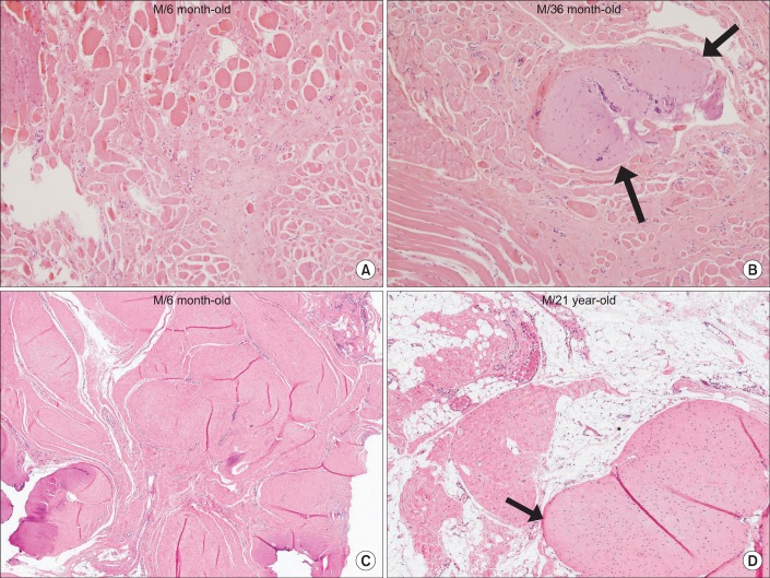
Fig. 7 The histopathological findings of the sternocleidomastoid muscle with congenital muscular torticollis. (A) Diffuse interstitial fibrosis with accompanying atrophic muscle fibers is noted (H&E, ×200). (B) Interstitial fibrosis with presence of aberrant tendon-like excessive dense connective tissue (arrows) (H&E, ×200). (C) Interstitial fibrosis with presence of aberrant tendon-like excessive dense connective tissue which was well-arranged (H&E, ×40). (D) Aberrant tendon-like excessive dense connective tissue and prominent fat infiltration (arrow) (H&E, ×40).
Cited by 5 articles
-
Congenital Muscular Torticollis Concurrent With Sagittal Synostosis: A Case Report
Seung-Hyun Kim, Ah-Reum Ahn, Shin-Young Yim
Ann Rehabil Med. 2014;38(5):712-716. doi: 10.5535/arm.2014.38.5.712.Quantitative Analysis of Magnetic Resonance Imaging of the Neck and Its Usefulness in Management of Congenital Muscular Torticollis
Jong Woo Kim, Seung Hyun Kim, Shin-Young Yim
Ann Rehabil Med. 2015;39(2):294-302. doi: 10.5535/arm.2015.39.2.294.Craniofacial Asymmetry in Adults With Neglected Congenital Muscular Torticollis
Kil-Yong Jeong, Kyung-Jay Min, Jieun Woo, Shin-Young Yim
Ann Rehabil Med. 2015;39(3):440-450. doi: 10.5535/arm.2015.39.3.440.Effectiveness of Surgical Release in Patients With Neglected Congenital Muscular Torticollis According to Age at the Time of Surgery
Kyung-Jay Min, Ah-Reum Ahn, Eun-Ji Park, Shin-Young Yim
Ann Rehabil Med. 2016;40(1):34-42. doi: 10.5535/arm.2016.40.1.34.Rare Concurrence of Congenital Muscular Torticollis and a Malignant Tumor in the Same Sternocleidomastoid Muscle
Yul-Hyun Park, Chul-Ho Kim, Jang-Hee Kim, Jun-Eun Park, Shin-Young Yim
Ann Rehabil Med. 2018;42(1):189-194. doi: 10.5535/arm.2018.42.1.189.
Reference
-
1. Chen MM, Chang HC, Hsieh CF, Yen MF, Chen TH. Predictive model for congenital muscular torticollis: analysis of 1021 infants with sonography. Arch Phys Med Rehabil. 2005; 86:2199–2203. PMID: 16271571.
Article2. Morrison DL, MacEwen GD. Congenital muscular torticollis: observations regarding clinical findings, associated conditions, and results of treatment. J Pediatr Orthop. 1982; 2:500–505. PMID: 7161384.3. Yim SY, Lee IY, Park MC, Kim JH. Differential diagnosis and management of abnormal posture of the head and neck. J Korean Med Assoc. 2009; 52:705–718.
Article4. Cheng JC, Wong MW, Tang SP, Chen TM, Shum SL, Wong EM. Clinical determinants of the outcome of manual stretching in the treatment of congenital muscular torticollis in infants. A prospective study of eight hundred and twenty-one cases. J Bone Joint Surg Am. 2001; 83-A:679–668. PMID: 11379737.5. Ablin DS, Jain K, Howell L, West DC. Ultrasound and MR imaging of fibromatosis colli (sternomastoid tumor of infancy). Pediatr Radiol. 1998; 28:230–233. PMID: 9545475.
Article6. Brans J, Aramideh M, Bosch A, Speelman H. Late presentation of congenital muscular torticollis: a non-dystonic cause of torticollis. J Neurol. 1996; 243:354–356. PMID: 8965110.
Article7. Entel RJ, Carolan FJ. Congenital muscular torticollis: magnetic resonance imaging and ultrasound diagnosis. J Neuroimaging. 1997; 7:128–130. PMID: 9128457.
Article8. Hayashi S, Ito K, Kogure T, Shimada M, Tsubuku M, Kaneko I, Kusama K. Clinical evaluation of congenital muscular torticollis by using MR imaging. Nihon Igaku Hoshasen Gakkai Zasshi. 1995; 55:957–960. PMID: 8570391.9. Parikh SN, Crawford AH, Choudhury S. Magnetic resonance imaging in the evaluation of infantile torticollis. Orthopedics. 2004; 27:509–515. PMID: 15181949.
Article10. Pereira S, Tani E, Skoog L. Diagnosis of fibromatosis colli by fine needle aspiration (FNA) cytology. Cytopathology. 1999; 10:25–29. PMID: 10068884.
Article11. Singer C, Green BA, Bruce JH, Bowen BC, Weiner WJ. Late presentation of congenital muscular torticollis: use of MR imaging and CT scan in diagnosis. Mov Disord. 1994; 9:100–103. PMID: 8139587.
Article12. Whyte AM, Lufkin RB, Bredenkamp J, Hoover L. Sternocleidomastoid fibrosis in congenital muscular torticollis: MR appearance. J Comput Assist Tomogr. 1989; 13:163–164. PMID: 2910939.13. Apple SK, Nieberg RK, Hirschowitz SL. Fine needle aspiration diagnosis of fibromatosis colli. A report of three cases. Acta Cytol. 1997; 41:1373–1376. PMID: 9990278.14. Kurtycz DF, Logrono R, Hoerl HD, Heatley DG. Diagnosis of fibromatosis colli by fine-needle aspiration. Diagn Cytopathol. 2000; 23:338–342. PMID: 11074630.
Article15. Nayak SP, Munshi MM, Bobhate SK. Cytodiagnosis of fibromatosis colli. Cytopathology. 2007; 18:266–268. PMID: 17635166.
Article16. Park IS, Kim L, Choi SJ, Han JY, Kim JM, Chu YC, Choi SG. Fine needle aspiration cytologic findings of fibromatosis colli: a report of three cases. Korean J Cytopathol. 2005; 16:61–65.17. Sauer T, Selmer L, Freng A. Cytologic features of fibromatosis colli of infancy. Acta Cytol. 1997; 41:633–635. PMID: 9167675.
Article18. Schwartz RA, Powers CN, Wakely PE Jr, Kellman RM. Fibromatosis colli. The utility of fine-needle aspiration in diagnosis. Arch Otolaryngol Head Neck Surg. 1997; 123:301–304. PMID: 9076237.
Article19. Cheng JC, Metreweli C, Chen TM, Tang S. Correlation of ultrasonographic imaging of congenital muscular torticollis with clinical assessment in infants. Ultrasound Med Biol. 2000; 26:1237–1241. PMID: 11120359.
Article20. Gonzales J, Ljung BM, Guerry T, Schoenrock LD. Congenital torticollis: evaluation by fine-needle aspiration biopsy. Laryngoscope. 1989; 99:651–654. PMID: 2725162.
- Full Text Links
-
- Actions
-
Cited
- CITED
-
- Close
- Share
-
- Similar articles
-
- Familial Congenital Muscular Torticollis: A Case Report
- Unipolar Release of the Sternocleidomastoideus in Congenital Muscular Torticollis in Children
- Quantitative Analysis of Magnetic Resonance Imaging of the Neck and Its Usefulness in Management of Congenital Muscular Torticollis
- Two Cases of Sternomastoid Tumor
- Biterminal Sternocleidomastoid Tenotomy for the Treatment of Congenital Muscular Torticollis in Children
