Injectate Volumes Needed to Reach Specific Landmarks and Contrast Pattern in Kambin's Triangle Approach with Spinal Stenosis
- Affiliations
-
- 1Department of Rehabilitation Medicine, Gachon University of Medicine and Science, Gil Medical Center, Incheon 405-760, Korea.
- 2Department of Radiology, Sanggye Paik Hospital, Inje University College of Medicine, Seoul 139-707, Korea.
- 3Department of Rehabilitation Medicine, Sanggye Paik Hospital, Inje University College of Medicine, Seoul 139-707, Korea. swc328@naver.com
- KMID: 2266718
- DOI: http://doi.org/10.5535/arm.2012.36.4.480
Abstract
OBJECTIVE
To identify the volumes of contrast material needed to reach the specific landmarks and contrast pattern during Kambin's triangle approach (KB-A) in lumbar spinal stenosis. METHOD: Sixty patients undergoing KB-A were investigated. Fifty-six patients were included in this study. KB-A were performed with the use of contrast-enhanced fluoroscopic visualization. After confirming the appropriate spinal needle position, a slow injection of up to 5.0 ml of nonionic contrast material was carried out. Under intermittent fluoroscopic guidance, contrast volumes were recorded as flow reached specific anatomic landmarks: ipsilateral inferior or superior neural foramen.
RESULTS
After 2.0 ml of contrast was injected, 93.2% of KB-A cases spread to the medial aspect of the inferior pedicle of the corresponding level of injection and 86.3% of KB-A spread to the medial aspect of the superior pedicle of the corresponding level of injection. After 3 ml of contrast was injected, 95.3% of KB-A spread to cover both the medial aspect of the inferior pedicle and the superior pedicle of the corresponding level of injection. A volume of 2 ml of injectate reaches the anterior epidural space 100% of the time.
CONCLUSION
This study demonstrates injectate volumes needed to reach the specific anatomic landmarks in KB-A. A volume of 3.0 ml of injectate reaches both the medial aspect of theinferior pedicle and the superior pedicle 94.6% of the time. Therefore, Interventionalists may consider a 1-level instead of a 2-level injection for patients with a bleeding risk or for 2 level central pathology.
Keyword
Figure
-
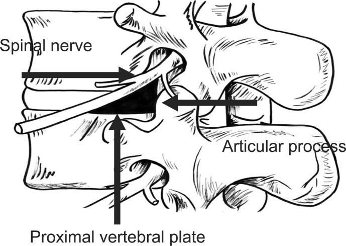
Fig. 1 Schematic description of the Kambin's triangle. The hypotenuse is the exiting nerve; the base is the caudad vertebral body; and the height is the traversing nerve root.
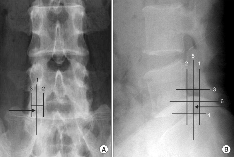
Fig. 2 (A) The anterior-posterior view of the lumbar spine, with the superimposed line (1) bisecting the pedicle. This line was drawn halfway between the farthest medial (2) and the farthest lateral (3) points on the pedicle. (B) The lateral view of the lumbar spine with the quadrant system superimposed. A line was drawn tangent to the curve of the spine at the level of interest along the posterior vertebral line. (1) A second line (2) was drawn parallel to the third line at the posterior margin of the foramen. Next, two lines perpendicular to lines 1 and 2 were drawn at the superior and inferior margins of the foramen (lines 3 and 4, respectively). Finally, line 5 was drawn bisecting lines 1 and 2, and line 6 was drawn bisecting lines 3 and 4. These lines divided the foramen into four quadrants. Arrow: needle position.

Fig. 3 (A) The anteroposterior view of a needle placed for a Kambin's triangle approach of the L5 nerve root demonstrating the landmarks used for this investigation. Note the contrast flowing along the most medial aspect of the inferior pedicle (PED). (B) The lateral view of a needle placed for a Kambin's triangle approach of the L5 nerve root demonstrating the landmarks used for this investigation. Note the contrast flowing anterior epidural space.
Reference
-
1. Slipman CW, Chow DW. Therapeutic spinal corticosteroid injections for the management of radiculopathies. Phys Med Rehabil Clin N Am. 2002; 13:697–711. PMID: 12380554.
Article2. Vad VB, Bhat AL, Lutz GE, Cammisa F. Transforaminal epidural steroid injections in lumbosacral radiculopathy: a prospective randomized study. Spine. 2002; 27:11–16. PMID: 11805628.3. Manchikanti L, Cash KA, Pampati V, Damron KS, McManus CD. Evaluation of lumbar transforaminal epidural injections with needle placement and contrast flow patterns: a prospective, descriptive report. Pain Physician. 2004; 7:217–223. PMID: 16868595.4. Murthy NS, Maus TP, Behrns CL. Intraforaminal location of the great anterior radiculomedullary artery (artery of Adamkiewicz): a retrospective review. Pain Med. 2010; 11:1756–1764. PMID: 21134118.
Article5. Botwin KP, Gruber RD, Bouchlas CG, Torres-Ramos FM, Sanelli JT, Freeman ED, Slaten WK, Rao S. Fluoroscopically guided lumbar transforaminal epidural steroid injections in degenerative lumbar stenosis: an outcome study. Am J Phys Med Rehabil. 2002; 81:898–905. PMID: 12447088.6. Kambin P, Brager MD. Percutaneous posterolateral discectomy. Anatomy and mechanism. Clin Orthop Relat Res. 1987; 223:145–154. PMID: 3652568.7. Kambin P. Arthroscopic microdiskectomy. Mt Sinai J Med. 1991; 58:159–164. PMID: 1857361.8. Furman MB, Mehta AR, Kim RE, Simon JI, Patel R, Lee TS, Reeves RS. Injectate volumes needed to reach specific landmarks in lumbar transforaminal epidural injections. PM R. 2010; 2:625–635. PMID: 20659718.
Article9. Hogan QH. Epidural anatomy examined by cryomicrotome section. Influence of age, vertebral level, and disease. Reg Anesth. 1996; 21:395–406. PMID: 8895998.10. Glaser SE, Shah RV. Root cause analysis of paraplegia following transforaminal epidural steroid injections: the 'unsafe' triangle. Pain Physician. 2010; 13:237–244. PMID: 20495587.11. Kambin P, Savitz MH. Arthroscopic microdiscectomy: an alternative to open disc surgery. Mt Sinai J Med. 2000; 67:283–287. PMID: 11021778.12. Jasper JF. Lumbar retrodiscal transforaminal injection. Pain Physician. 2007; 10:501–510. PMID: 17525785.
Article13. Lew HL, Coelho P, Chou LH. Preganglionic approach to transforaminal epidural steroid injections. Am J Phys Med Rehabil. 2004; 83:378. PMID: 15100628.
Article14. Lee IS, Kim SH, Lee JW, Hong SH, Choi JY, Kang HS, Song JW, Kwon AK. Comparison of the temporary diagnostic relief of transforaminal epidural steroid injection approaches: conventional versus posterolateral technique. AJNR Am J Neuroradiol. 2007; 28:204–208. PMID: 17296980.
- Full Text Links
-
- Actions
-
Cited
- CITED
-
- Close
- Share
-
- Similar articles
-
- Kambin's Triangle Approach of Lumbar Transforaminal Epidural Injection with Spinal Stenosis
- Full-endoscopic Trans-Kambin’s Triangle Lumbar Interbody Fusion: Technique and Review of Literature
- Get Ready for 100 Years of Active Spine Life Using Percutaneous Endoscopic Spine Surgery (PESS)
- Anatomic Considerations of Intervertebral Disc Perspective in Lumbar Posterolateral Approach via Kambin's Triangle: Cadaveric Study
- Cadaveric anatomy of the lumbar triangular safe zone of Kambin’s in North West Indian population
