Spontaneous Perirenal Hemorrhage in Cauda Equina Syndrome: A Case Report
- Affiliations
-
- 1Department of Physical Medicine and Rehabilitation, Soonchunhyang University College of Medicine, Bucheon, Korea. pmrguren@gmail.com
- KMID: 2266620
- DOI: http://doi.org/10.5535/arm.2013.37.4.595
Abstract
- Neurogenic bladder is a common cause of acute pyelonephritis (APN) in cauda equina syndrome (CES). Perirenal hemorrhage, a rare complication of APN, can be a life-threatening condition. To our knowledge, there is no previous report of perirenal hemorrhage as a complication of APN in CES. A 57-year-old male, diagnosed with CES, due to a L3 burst fracture 3 months earlier, was presented with fever and chills. His diagnosis was APN due to neurogenic bladder. After treatment for APN, he was transferred to the department of rehabilitation medicine for management of his CES. Because of large post-voiding residual urine volumes, he performed self-catheterization after voiding. However, he presented again with fever and chills, and recurrent APN was diagnosed. On the third day of antibiotic treatment, he had acute abdominal pains and hypovolemic shock. Abdominal computed tomography and angiography showed left APN and a perirenal hematoma with left renal capsular artery bleeding. After embolization of the left renal capsular artery, no further active bleeding occurred. Because APN due to neurogenic bladder can lead to critical complications, such as perirenal hemorrhage, the physician should pay attention to the early diagnosis and treatment of urinary tract infection and the management of neurogenic bladder after CES.
MeSH Terms
Figure
-
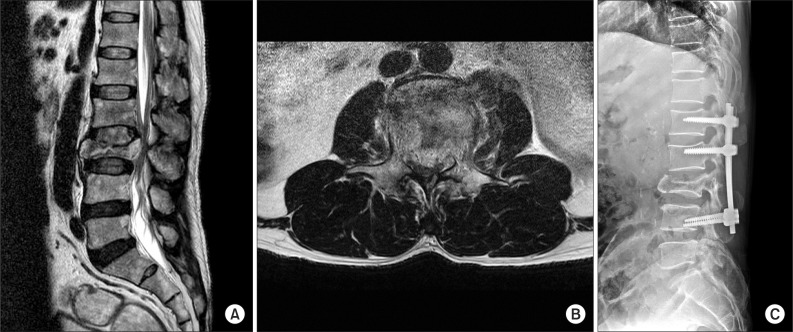
Fig. 1 Initial T2-weighted magnetic resonance imaging scans show (A) burst fracture at L3 vertebral body and (B) retro-pulsed bony fragments into spinal canal. (C) Postoperation X-ray demonstrates posterior fixation state in L1-4.
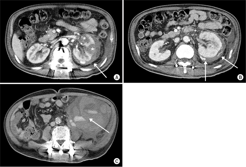
Fig. 2 Contrast enhanced abdominal computerized tomography shows (A) multiple hypoattenuation of left kidney parenchyme (asterisk) and hypoattenuated fluid collection with density of blood in the perinephric space (arrow), (B) active extravasation of contrast media at the left renal capsular artery (arrow), and (C) hypoattenuated fluid collection with contrast leakage in lower left quadrant pelvic cavity (arrow).
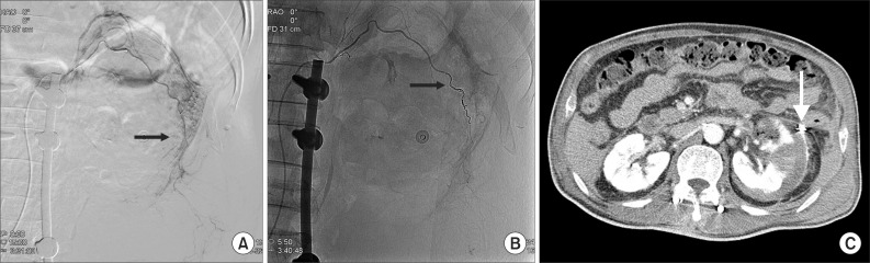
Fig. 3 Selective left renal angiography shows suspicious contrast leakage of left renal capsular artery (A, arrow) and no more contrast leakage after renal capsular artery embolization with microcoils (B, arrow). Follow-up abdominal computerized tomography shows no visualization of active bleeding and embolization state with microcoils in left renal capsular artery (C, arrow).
Reference
-
1. Gitelman A, Hishmeh S, Morelli BN, Joseph SA Jr, Casden A, Kuflik P, et al. Cauda equina syndrome: a comprehensive review. Am J Orthop (Belle Mead NJ). 2008; 37:556–562. PMID: 19104682.2. Shin JC, Park CI, Rha DW, Chon J, Kim JE, Jeon SC, et al. The diagnosis of upper urinary tract infection using abdominal computerized tomography in spinal cord injured patients. J Korean Acad Rehabil Med. 2004; 28:140–145.3. Song HJ, Choi MY, Kim MY, Lee YS, Kim SJ, Choi GB, et al. A case of spontaneous rupture of the kidney with acute pyelonephritis. Korean J Nephrol. 2003; 22:757–762.4. Chang TH, Wu WJ, Hsiao HL, Yeh HC, Huang CH, Lee YC. Spontaneous perirenal hematoma: a case report. Kaohsiung J Med Sci. 2005; 21:578–581. PMID: 16670051.
Article5. McDougal WS, Kursh ED, Persky L. Spontaneous rupture of the kidney with perirenal hematoma. J Urol. 1975; 114:181–184. PMID: 1159905.
Article6. Albi G, del Campo L, Tagarro D. Wunderlich's syndrome: causes, diagnosis and radiological management. Clin Radiol. 2002; 57:840–845. PMID: 12384111.7. Yoon JC, Kim W, Cho GC, Hong JS, Lee MW, Jang SE, et al. Idiopathic spontaneous renal rupture. J Korean Soc Emerg Med. 2001; 12:523–527.8. Aizenstein RI, Owens C, Sabnis S, Wilbur AC, Hibbeln JF, O'Neil HK. The perinephric space and renal fascia: review of normal anatomy, pathology, and pathways of disease spread. Crit Rev Diagn Imaging. 1997; 38:325–367. PMID: 9376088.9. Polkey HJ, Vynalek WJ. Spontaneous nontraumatic perirenal and renal hematomas: an experimental and clinical study. Arch Surg. 1933; 26:196–218.10. Bryce TN, Ragnarsson KT, Stein AB. Spinal cord injury. In : Braddom RL, editor. Physical medicine and rehabilitation. 3rd ed. Philadephia: Sauders;2007. p. 1328–1329.
- Full Text Links
-
- Actions
-
Cited
- CITED
-
- Close
- Share
-
- Similar articles
-
- Cavernous Hemangioma of the Cauda Equina
- A Case of Myxopapillary Ependymoma in the Cauda Equina: Case Report
- Causes and Clinical Manifestations of Cauda Equina Syndrome
- Cauda Equina Syndrome after Spinal Manipulative Therapy: A case report
- Hypertrophic Interstitial Neuritis of Cauda Equina: Case Report with Myelographic Findings
