Ann Dermatol.
2012 Nov;24(4):468-471. 10.5021/ad.2012.24.4.468.
Dermoscopy: A Useful Tool for the Diagnosis of Angiokeratoma
- Affiliations
-
- 1Department of Dermatology, Gangnam Severance Hospital, Yonsei University College of Medicine, Seoul, Korea. ydshderm@yuhs.ac
- 2Department of Dermatology, Severance Hospital, Cutaneous Biology Research Institute, Yonsei University College of Medicine, Seoul, Korea.
- 3Department of Dermatology, Bundang CHA Medical Center, CHA University, Seongnam, Korea.
- KMID: 2266045
- DOI: http://doi.org/10.5021/ad.2012.24.4.468
Abstract
- Angiokeratoma is a rare vascular malformation of the upper dermis that presents clinically as deep red to blue-black in color and tends to take a diverse configuration without self-limiting. Here, we reported dermoscopic findings by two cases of angiokeratoma; solitary angiokeratoma and angiokeratoma circumscriptum. A 24-year-old male presented with a 2-months history of 5 mm sized black colored papule on the right buttock. A dermoscopic pattern characterized by red and dark lacunae, whitish veil covered with scale. A 26-year-old woman presented with multiple, 2~10 mm, dark colored papules on the anterior neck with zosteriform fashion since childhood. A dermoscopic pattern presented by red lacunae intermingled with whitish veil. As a previous report, our two cases was the most common dermoscopic pattern of angiokeratoma; red lacunae and whitish veil. Angiokeratoma is often diagnosed as melanocytic nevi, Spitz nevi, malignant melanomas, pigmented basal cell carcinomas, seborrheic keratoses, dermatofibromas and other vascular lesions including hemangiomas and pyogenic granulomas. However, in the dermoscopic view, these above lesions hardly show red lacunae with whitish veils. Therefore, the dermscopic view is a useful differential method of angiokeratoma.
Keyword
MeSH Terms
Figure
-
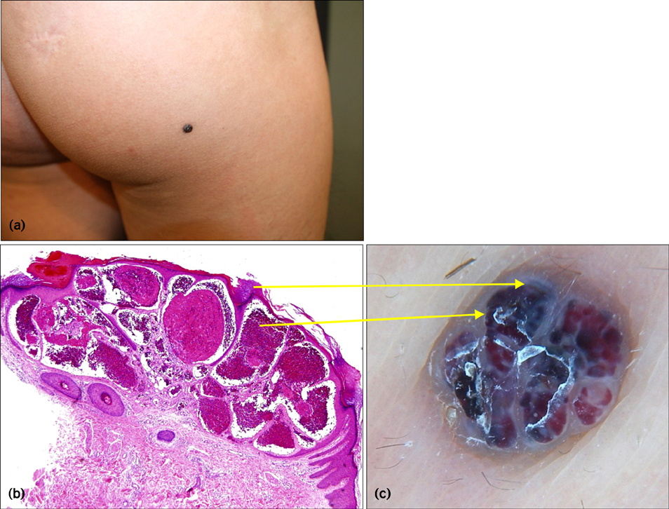
Fig. 1 (a) Five-millimeter black lesion is shown on the right buttock. (b) Histopathology and (c) dermoscopy of the lesion are shown. Dilated vascular spaces in the papillary dermis with thrombosis presented as dark lacunae. Hyperkeratosis and acanthosis appeared as a whitish veil (H&E, ×100).

Fig. 2 (a) Multiple dark zosteriform papules on the anterior neck, shown macroscopically. (b) Dermoscopy of the lesion showed red and black lacunae with a whitish veil. (c) Microscopic appearance of the lesion showing hyperkeratosis and several dermal dilated vessels fully congested with red blood cells (H&E, ×100).
Reference
-
1. Wolf IH. Dermoscopic diagnosis of vascular lesions. Clin Dermatol. 2002. 20:273–275.
Article2. Zaballos P, Daufí C, Puig S, Argenziano G, Moreno-Ramírez D, Cabo H, et al. Dermoscopy of solitary angiokeratomas: a morphological study. Arch Dermatol. 2007. 143:318–325.3. Sahin MT, Türel-Ermertcan A, Oztürkcan S, Türkdogan P. Thrombosed solitary angiokeratoma of Mibelli simulating malignant melanoma: the importance of dermoscopy in differential diagnosis. J Eur Acad Dermatol Venereol. 2006. 20:102–104.4. Mittal R, Aggarwal A, Srivastava G. Angiokeratoma circumscriptum: a case report and review of the literature. Int J Dermatol. 2005. 44:1031–1034.
Article5. Naranjo Sintes R, Pereda Hernandez J, Delgado Florencio V, Linares Solano J. Angiokeratoma. Apropos of 93 cases. Med Cutan Ibero Lat Am. 1988. 16:255–261.6. Campos-do-Carmo G, Ramos-e-Silva M. Dermoscopy: basic concepts. Int J Dermatol. 2008. 47:712–719.
Article7. Zaballos P, Llambrich A, Cuéllar F, Puig S, Malvehy J. Dermoscopic findings in pyogenic granuloma. Br J Dermatol. 2006. 154:1108–1111.
Article8. Sakakibara A, Kamijima M, Shibata S, Yasue S, Kono M, Tomita Y. Dermoscopic evaluation of vascular structures of various skin tumors in Japanese patients. J Dermatol. 2010. 37:316–322.
Article
- Full Text Links
-
- Actions
-
Cited
- CITED
-
- Close
- Share
-
- Similar articles
-
- Role of dermoscopy and biopsy in the diagnosis of skin cancer: it takes two to tango
- Xanthogranuloma for Whom Dermoscopy Was Used as an Adjuvant Diagnostic Tool
- A Case of Angiokeratoma of the Vulva
- The Diagnostic Accuracy of Dermoscopy for Scabies
- Successfully Treated Basal Cell Carcinoma with Mohs Surgery, Diagnosing with Dermoscopy as a Primary Diagnostic Tool without Preoperative Punch Biopsy: A Report of Two Cases
