Ann Dermatol.
2013 Aug;25(3):385-386. 10.5021/ad.2013.25.3.385.
Primary Dermal Melanoma Latent for More than 10 Years
- Affiliations
-
- 1Department of Dermatology, Faculty of Medicine, Kagawa University, Kagawa, Japan. junjun@med.kagawa-u.ac.jp
- 2Department of Diagnostic Pathology, Faculty of Medicine, Kagawa University, Kagawa, Japan.
- 3Department of Dermatology, Takamatsu Red Cross Hospital, Kagawa, Japan.
- KMID: 2265888
- DOI: http://doi.org/10.5021/ad.2013.25.3.385
Abstract
- No abstract available.
MeSH Terms
Figure
-
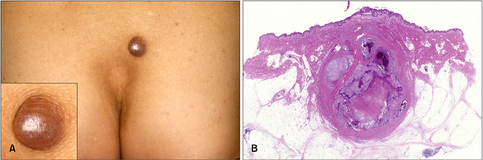
Fig. 1 (A) Brown/red nodule on the sacral region. (B) Primary dermal melanoma composed of a dermal and subcutaneous nodule surrounded by fibrous tissue, with extensive central necrosis and hemorrhaging (H&E, ×10).

Fig. 2 Immunohistochemical findings of primary dermal melanoma. (A) HMB-45 staining was focally positive (×200). (B) Ki-67 staining was low positive (14.9% of cells were positive) (×200).
Reference
-
1. Bowen GM, Chang AE, Lowe L, Hamilton T, Patel R, Johnson TM. Solitary melanoma confined to the dermal and/or subcutaneous tissue: evidence for revisiting the staging classification. Arch Dermatol. 2000; 136:1397–1399.2. Cassarino DS, Cabral ES, Kartha RV, Swetter SM. Primary dermal melanoma: distinct immunohistochemical findings and clinical outcome compared with nodular and metastatic melanoma. Arch Dermatol. 2008; 144:49–56.3. Balch CM, Gershenwald JE, Soong SJ, Thompson JF, Atkins MB, Byrd DR, et al. Final version of 2009 AJCC melanoma staging and classification. J Clin Oncol. 2009; 27:6199–6206.
Article4. Hida Y, Kubo Y, Miyajima O, Arase S. Primary dermal melanoma: a case report and molecular characterization. J Dermatol. 2009; 36:346–352.
Article
- Full Text Links
-
- Actions
-
Cited
- CITED
-
- Close
- Share
-
- Similar articles
-
- Primary malignant melanoma arising in a cystic teratoma
- A Case of Primary Malignant Melanoma of the Vagina: Trial of a Wide Local Excision of Vagina and Rectum
- Recurrent Primary Meningeal Melanoma: Case Report
- Primary Malignant Melanoma of the Cervical Spinal Nerve Root: A Case Report
- Immunohistochemical Study on the Expression of Mutated p53 Protein and Bcl-2 Protein in Melanocytic Lesions of Skin
