Generalized Dowling-Degos Disease: Case Reports
- Affiliations
-
- 1Division of Dermatology, Department of Medicine, Faculty of Medicine, Chulalongkorn University and King Chulalongkorn Memorial Hosptial, Thai Red Cross Society, Bangkok, Thailand. jademdcu@yahoo.com
- KMID: 2265879
- DOI: http://doi.org/10.5021/ad.2013.25.3.360
Abstract
- Dowling-Degos disease (DDD) is a rare autosomal dominant trait characterized by numerous, symmetrical, progressive and pigmented macules over the axillae, groins, face, neck, arms and trunk as well as scattered comedo-like lesions (dark dot, follicles) and pitted acneiform scars. Histopathology is diagnostic testing using a distinctive form of acanthosis, characterized by an irregular elongation of thin branching rete ridges, with a concentration of melanin at the tips. We report cases of generalized DDD in a single family with autosomal dominant penetrance. DDD can be presented in a generalized form with hypopigmented lesions instead of reticulate hyperpigmentation confined to the flexor areas. This form can be differentiated from DUH by histopathology.
MeSH Terms
Figure
-
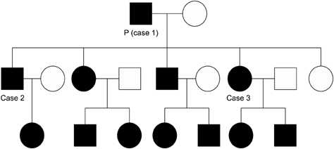
Fig. 1 Pedigree of family with generalized Dowling-Degos disease (DDD). Proband (P), his first son (case 2), and his second daughter (case 3) had generalized DDD.
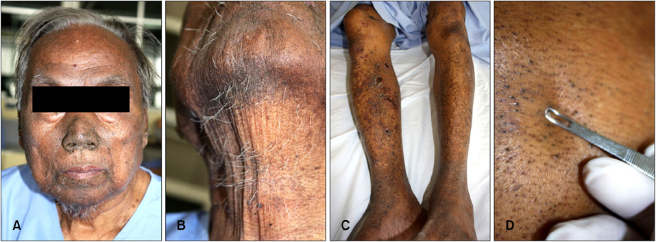
Fig. 2 Skin lesion of proband, 82-year-old man. (A, B) Reticulate hyperpigmentation, multiple pits and comedo-like lesions distributed over the face, perioral area and neck. (C) Reticulate hyperpigmentation with symmetrically distributed hypopigmented macules on shins. (D) Comedo-like lesions distributed over the back.

Fig. 3 Son of proband, 58-year-old man. (A) Reticulate pigmented macules, multiple pits and comedo-like lesions distributed over the face, perioral area and neck. (B) Reticulate hyperpigmentation on axilla.

Fig. 4 Daughter of proband, 50-year-old woman. (A) Reticulate pigmented macules, multiple pits and comedo-like lesions distributed over the face, perioral area and neck. (B, C) Reticulate hyperpigmentation on forearms.

Fig. 5 Skin biopsy specimens from proband of hyperigmented macule (A) and comedo-like lesion (B). (A) Epidermal hyperkeratosis, acanthosis, irregular elongated thin branching rete ridges growing down into the dermis and increased melanin pigment in the lower part of rete pegs (H&E, ×200). (B) Keratin-filled cysts resembling comedones with irregular elongated thin branching rete ridges growing down into the dermis and increased melanin pigment in the lower part of rete pegs (H&E, ×40).
Reference
-
1. Crovato F, Nazzari G, Rebora A. Dowling-Degos disease (reticulate pigmented anomaly of the flexures) is an autosomal dominant condition. Br J Dermatol. 1983; 108:473–476.
Article2. Rebora A, Crovato F. The spectrum of Dowling-Degos disease. Br J Dermatol. 1984; 110:627–630.
Article3. Kim YC, Davis MD, Schanbacher CF, Su WP. Dowling-Degos disease (reticulate pigmented anomaly of the flexures): a clinical and histopathologic study of 6 cases. J Am Acad Dermatol. 1999; 40:462–467.
Article4. Lestringant GG, Masouyé I, Frossard PM, Adeghate E, Galadari IH. Co-existence of leukoderma with features of Dowling-Degos disease: reticulate acropigmentation of Kitamura spectrum in five unrelated patients. Dermatology. 1997; 195:337–343.
Article5. Ostlere L, Holden CA. Dowling-Degos disease associated with Kitamura's reticulate acropigmentation. Clin Exp Dermatol. 1994; 19:492–495.
Article6. Sandhu K, Saraswat A, Kanwar AJ. Dowling-Degos disease with dyschromatosis universalis hereditaria-like pigmentation in a family. J Eur Acad Dermatol Venereol. 2004; 18:702–704.
Article7. Thami GP, Jaswal R, Kanwar AJ, Radotra BD, Singh IP. Overlap of reticulate acropigmentation of Kitamura, acropigmentation of Dohi and Dowling-Degos disease in four generations. Dermatology. 1998; 196:350–351.
Article8. Harper JI, Trembath RC. Genetics and genodermatoses. In : Rook AJ, Burns T, editors. Rook's textbook of dermatology. 7th ed. Malden, Mass: Blackwell Science;2004. p. 15.11–15.13.9. Howell JB, Freeman RG. Reticular pigmented anomaly of the flexures. Arch Dermatol. 1978; 114:400–403.
Article10. Betz RC, Planko L, Eigelshoven S, Hanneken S, Pasternack SM, Bussow H, et al. Loss-of-function mutations in the keratin 5 gene lead to Dowling-Degos disease. Am J Hum Genet. 2006; 78:510–519.
Article11. Li CR, Xing QH, Li M, Qin W, Yue XZ, Zhang XJ, et al. A gene locus responsible for reticulate pigmented anomaly of the flexures maps to chromosome 17p13.3. J Invest Dermatol. 2006; 126:1297–1301.
Article12. Crovato F, Desirello G, Rebora A. Is Dowling-Degos disease the same disease as Kitamura's reticulate acropigmentation? Br J Dermatol. 1983; 109:105–110.
Article13. Berth-Jones J, Graham-Brown RA. A family with Dowling Degos disease showing features of Kitamura's reticulate acropigmentation. Br J Dermatol. 1989; 120:463–466.
Article14. Cox NH, Long E. Dowling-Degos disease and Kitamura's reticulate acropigmentation: support for the concept of a single disease. Br J Dermatol. 1991; 125:169–171.
Article15. Al Hawsawi K, Al Aboud K, Alfadley A, Al Aboud D. Reticulate acropigmentation of Kitamura-Dowling Degos disease overlap: a case report. Int J Dermatol. 2002; 41:518–520.
Article16. Oyama M, Shimizu H, Ohata Y, Tajima S, Nishikawa T. Dyschromatosis symmetrica hereditaria (reticulate acropigmentation of Dohi): report of a Japanese family with the condition and a literature review of 185 cases. Br J Dermatol. 1999; 140:491–496.
Article17. Bonifas JM, Rothman AL, Epstein EH Jr. Epidermolysis bullosa simplex: evidence in two families for keratin gene abnormalities. Science. 1991; 254:1202–1205.
Article18. Bonifas JM, Bare JW, Lynch ED, Lebo RV, Epstein EH Jr. Regional assignment of the human keratin 5 (KRT5) gene to chromosome 12q near D12S14 by PCR analysis of somatic cell hybrids and multicolor in situ hybridization. Genomics. 1992; 13:452–454.
Article19. Stephens K, Ehrlich P, Weaver M, Le R, Spencer A, Sybert VP. Primers for exon-specific amplification of the KRT5 gene: identification of novel and recurrent mutations in epidermolysis bullosa simplex patients. J Invest Dermatol. 1997; 108:349–353.
Article20. Wu YH, Lin YC. Generalized Dowling-Degos disease. J Am Acad Dermatol. 2007; 57:327–334.
Article
- Full Text Links
-
- Actions
-
Cited
- CITED
-
- Close
- Share
-
- Similar articles
-
- A Case of Dowling-Degos Disease
- A Case of Reticulate Pigmented Anomaly of the Flexures (Dowling-Degos Disease)
- A Case of Dowling-degos Disease Affecting the Vulva
- A Case of Reticulate Pigmented Anomaly of the Flexures
- A case of reticulate acropigmentation of Kitamura with hyperpigmented macular lesions on the scrotum
