Ann Dermatol.
2014 Feb;26(1):123-124. 10.5021/ad.2014.26.1.123.
Superficial Acral Fibromyxoma on the Palm
- Affiliations
-
- 1Department of Dermatology, Samsung Medical Center, Sungkyunkwan University School of Medicine, Seoul, Korea. dylee@skku.edu
- KMID: 2265714
- DOI: http://doi.org/10.5021/ad.2014.26.1.123
Abstract
- No abstract available.
MeSH Terms
Figure
-
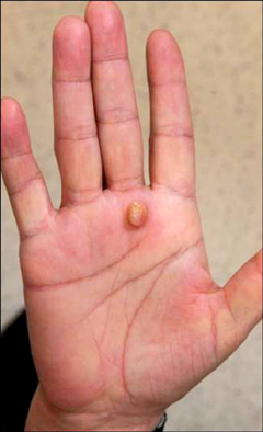
Fig. 1 A nodular mass on the palm side of right hand.

Fig. 2 Histopathological and immunohistochemical findings included (A) dome-shaped nodule (H&E, ×40), (B) moderate cellular proliferation of spindled fibroblast-like and stellate cells embedded in myxoid matrix (H&E, ×200), negative staining of tumor cells for CD34 (C: ×200) and epithelial membrane antigen (D: ×200), and positive staining for CD99 (E: ×200).
Reference
-
1. Fetsch JF, Laskin WB, Miettinen M. Superficial acral fibromyxoma: a clinicopathologic and immunohistochemical analysis of 37 cases of a distinctive soft tissue tumor with a predilection for the fingers and toes. Hum Pathol. 2001; 32:704–714.
Article2. Hollmann TJ, Bovée JV, Fletcher CD. Digital fibromyxoma (superficial acral fibromyxoma): a detailed characterization of 124 cases. Am J Surg Pathol. 2012; 36:789–798.3. Tardío JC, Butrón M, Martín-Fragueiro LM. Superficial acral fibromyxoma: report of 4 cases with CD10 expression and lipomatous component, two previously underrecognized features. Am J Dermatopathol. 2008; 30:431–435.
Article4. Goo J, Jung YJ, Kim JH, Lee SY, Ahn SK. A case of recurrent superficial acral fibromyxoma. Ann Dermatol. 2010; 22:110–113.
Article
- Full Text Links
-
- Actions
-
Cited
- CITED
-
- Close
- Share
-
- Similar articles
-
- A Case of Superficial Acral Fibromyxoma Showing Erythronychia
- A Case of Recurrent Superficial Acral Fibromyxoma
- A Case of Superficial Acral Fibromyxoma Occurring after Trauma in a Childhood Patient
- A Case of Superficial Acral Fibromyxoma
- Solitary Superficial Acral Fibromyxoma on the Foot: A Case Report and Brief Literature Review
