A Case of Recurrent Superficial Acral Fibromyxoma
- Affiliations
-
- 1Department of Dermatology, Wonju College of Medicine, Yonsei University, Wonju, Korea. ahnsk@wonju.yonsei.ac.kr
- KMID: 2172054
- DOI: http://doi.org/10.5021/ad.2010.22.1.110
Abstract
- Superficial acral fibromyxoma (SAFM) is a rare myxoid tumor that was first described in 2001. The presence of a very slow growing solitary tender mass in the subungual area is the typical clinical feature at presentation. Histopathologically, SAFM is composed of stellate cells in a myxocollagenous matrix with a poorly circumscribed margin. This tumor is thought to be benign, but its natural course is not fully understood. We describe a 15-year-old patient with recurrent SAFM and discuss the proper treatment and follow up.
Keyword
MeSH Terms
Figure
-

Fig. 1 The tender, growing subungual lesion and the distorted nail plate. The right side of the nail plate looked pale because of the pressing margin of the tumor.

Fig. 2 (A) Poorly circumscribed dermal tumor with myxoid and fibrous areas (H&E, ×40). (B) Stellate or fibroblast-like cells showing a fascicular growth pattern (H&E, ×100). (C) Stellate cells with an elongated thin cytoplasm were noted in the collagenous and mucinous areas. Blood vessels proliferated focally. (H&E, ×200).
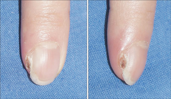
Fig. 3 A recurrent tumor was noted 1 year 8 months after the initial procedure.
Cited by 3 articles
-
Superficial Acral Fibromyxoma on the Palm
Se-Won Park, Jun-Hwan Kim, Hyun-Tae Shin, Ji Ho Park, Jong-Hee Lee, Dong-Youn Lee, Joo-Heung Lee, Jun-Mo Yang
Ann Dermatol. 2014;26(1):123-124. doi: 10.5021/ad.2014.26.1.123.Diagnostic Pitfalls of Differentiating Cellular Digital Fibroma from Superficial Acral Fibromyxoma
Jin Yong Lee, So Eun Park, Soo Jung Shin, Chul Woo Kim, Sang Seok Kim
Ann Dermatol. 2015;27(4):462-464. doi: 10.5021/ad.2015.27.4.462.Recurrent Huge Benign Tumors in the Hands
Min Wook Kim, So Min Hwang, Kwang Ryeol Lim, Yong Hui Jung, Jennifer K Song
J Korean Soc Surg Hand. 2012;17(4):153-158. doi: 10.12790/jkssh.2012.17.4.153.
Reference
-
1. Fetsch JF, Laskin WB, Miettinen M. Superficial acral fibromyxoma: a clinicopathologic and immunohistochemical analysis of 37 cases of a distinctive soft tissue tumor with a predilection for the fingers and toes. Hum Pathol. 2001. 32:704–714.
Article2. Kazakov DV, Mentzel T, Burg G, Kempf W. Superficial acral fibromyxoma: report of two cases. Dermatology. 2002. 205:285–288.
Article3. Andre J, Theunis A, Richert B, de Saint-Aubain N. Superficial acral fibromyxoma: clinical and pathological features. Am J Dermatopathol. 2004. 26:472–474.
Article4. Meyerle JH, Keller RA, Krivda SJ. Superficial acral fibromyxoma of the index finger. J Am Acad Dermatol. 2004. 50:134–136.
Article5. Quaba O, Evans A, Al-Nafussi AA, Nassan A. Superficial acral fibromyxoma. Br J Plast Surg. 2005. 58:561–564.
Article6. Abou-Nukta F, Fiedler P, Parkash V, Arons J. Superficial acral fibromyxoma of the distal phalanx of the thumb. J Hand Surg Br. 2006. 31:619–620.
Article7. Oteo-Alvaro A, Meizoso T, Scarpellini A, Ballestin C, Perez-Espejo G. Superficial acral fibromyxoma of the toe, with erosion of the distal phalanx. A clinical report. Arch Orthop Trauma Surg. 2008. 128:271–274.
Article8. Frierson HF, Cooper PH. Myxoid variant of dermatofibrosarcoma protuberans. Am J Surg Pathol. 1983. 7:445–450.
Article9. Graadt van Roggen JF, Hogendoorn PC, Fletcher CD. Myxoid tumours of soft tissue. Histopathology. 1999. 35:291–312.
Article10. Zelger BG, Calonje E, Zelger B. Myxoid dermatofibroma. Histopathology. 1999. 34:357–364.
Article
- Full Text Links
-
- Actions
-
Cited
- CITED
-
- Close
- Share
-
- Similar articles
-
- A Case of Superficial Acral Fibromyxoma Showing Erythronychia
- A Case of Superficial Acral Fibromyxoma Occurring after Trauma in a Childhood Patient
- A Case of Superficial Acral Fibromyxoma
- Solitary Superficial Acral Fibromyxoma on the Foot: A Case Report and Brief Literature Review
- Superficial Acral Fibromyxoma on the Second Toe
