Ann Dermatol.
2014 Aug;26(4):552-554. 10.5021/ad.2014.26.4.552.
An Unusual Presentation of a Progressive Zosteriform Macular Pigmented Lesion
- Affiliations
-
- 1Department of Dermatology, College of Medicine, The Catholic University of Korea, Seoul, Korea. cjpark777@yahoo.co.kr
- KMID: 2265613
- DOI: http://doi.org/10.5021/ad.2014.26.4.552
Abstract
- No abstract available.
Figure
-

Fig. 1 Linear macular pigmented lesion extending from right to left, across the midline of the trunk.
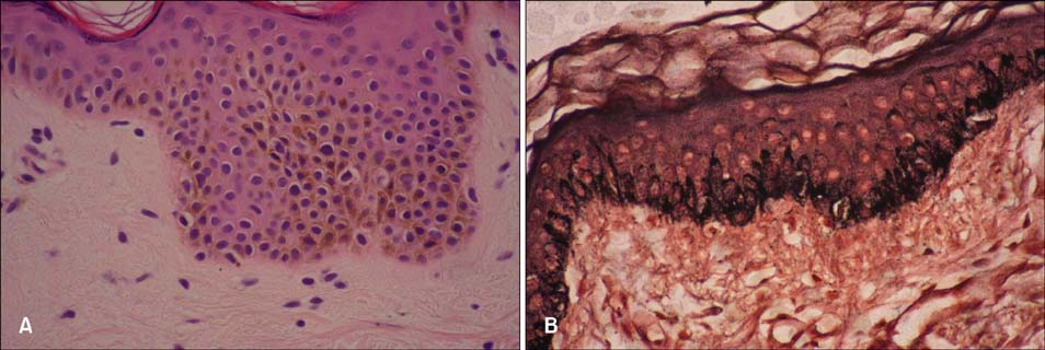
Fig. 2 (A) Increased pigmentation in the basal layer and some melanophages in the upper dermis (H&E, ×400), (B) Immunohistochemical staining is positive for Fontana-Masson (×200).
Reference
-
1. Rower JM, Carr RD, Lowney ED. Progressive cribriform and zosteriform hyperpigmentation. Arch Dermatol. 1978; 114:98–99.
Article2. Simões GA, Piva N. Progressive zosteriform macular pigmented lesions. Arch Dermatol. 1980; 116:20.
Article3. Lee JH, Sung KJ, Kim WS. A case of progressive cribriform and zosteriform hyperpigmentation. Korean J Dermatol. 1981; 19:515–519.
- Full Text Links
-
- Actions
-
Cited
- CITED
-
- Close
- Share
-
- Similar articles
-
- A Case of Progressive Zosteriform Macular Pigmented Lesion
- A Case of Progressive Zosteriform Macular Pigmented Lesion
- Progressive Zosteriform Macular Pigmented Lesion
- A Case of Progressive Cribriform and Zosteriform Hyperpigmentation
- A Case of Atypical Progressive Cribriform and Zosteriform Hyperpigmentation
