Three Cases of Lichen Nitidus Associated with Various Cutaneous Diseases
- Affiliations
-
- 1Department of Dermatology, Hallym University Sacred Heart Hospital, Anyang, Korea. dermakkh@naver.com
- KMID: 2265594
- DOI: http://doi.org/10.5021/ad.2014.26.4.505
Abstract
- Lichen nitidus (LN) is an uncommon, usually asymptomatic cutaneous eruption characterized by the presence of multiple, small, flesh-colored papules. The epidemiologic and pathophysiologic characteristics of LN have not yet been defined. Furthermore, LN has rarely been described in association with other cutaneous diseases. We herein report 3 cases of LN associated with various cutaneous diseases, including lichen striatus, oral lichen planus, and psoriasis vulgaris.
Keyword
Figure
-
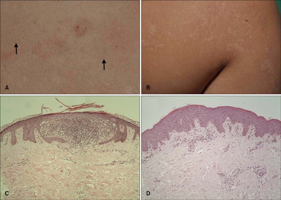
Fig. 1 Physical examination of the patient 1 revealed well-defined, pinhead-sized, skin-colored to erythematous grouped papules (arrows) on the right upper back (A), and pinhead-sized, whitish papules in a linear configuration on the right arm (B). Histopathologic examination from the right upper back shows hyperkeratosis, acanthosis, hydropic degeneration of the basal cell layer, and 'claw clutching a ball' appearance (C), from the the right arm shows hyperkeratosis, focal parakeratosis, acanthosis, hydropic degeneration of the basal cell layer, superficial perivascular lymphocytic infiltration and perifollicular lymphohistiocytic infiltration (D) (C, D: H&E, ×100).
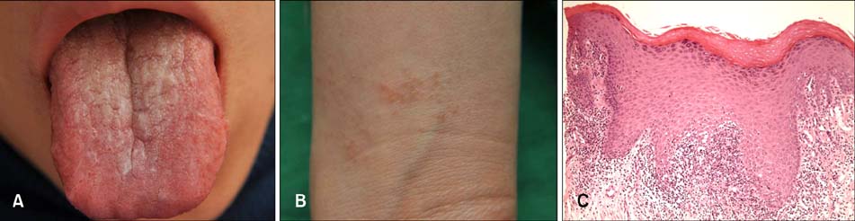
Fig. 2 Physical examination of the patient 2 revealed slightly erythematous plaques with whitish striae arranged lacy pattern on the dorsal surface on the tongue (A), and localized, relatively well defined, 2 to 3 mm sized, skin colored to slightly erythematous, grouped papules on both wrists (B). (C) Biopsy from the tongue shows hyperkeratosis, hypergranulosis, irregular acanthosis, hydropic degeneration of basal cells and band like infiltration of lymphocytes in the upper dermis (H&E, ×100).
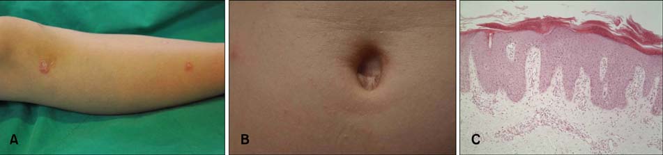
Fig. 3 Physical examination of the patient 3 revealed scattered, relatively well-defined, erythematous, scaly plaques on the lower extremities (A), and scattered, well-defined, pinhead-sized, skin colored, glistening papules on the trunk (B). (C) Biopsy from the right knee shows parakeratotic mounds (Munro microabscesses), hyperkeratosis, acanthosis with elongation of rete ridges and dilated capillaries at the superficial dermis with perivascular lymphocytic infiltration (H&E, ×100).
Reference
-
1. Pinkus F. Verhand. Berlin Dermat Gesel. 1901; 12:3.2. Park JH, Kye YC, Kim SN. A clinical and histopathologic study of lichen nitidus. Korean J Dermatol. 2003; 41:857–868.3. Munro CS, Cox NH, Marks JM, Natarajan S. Lichen nitidus presenting as palmoplantar hyperkeratosis and nail dystrophy. Clin Exp Dermatol. 1993; 18:381–383.
Article4. Thibaudeau A, Maillard H, Croué A, Belperron P, Avenel Audran M, Verret JL. Palmoplantar lichen nitidus: a rare cause of palmoplantar hyperkeratosis. Ann Dermatol Venereol. 2004; 131:822–824.5. Natarajan S, Dick DC. Lichen nitidus associated with nail changes. Int J Dermatol. 1986; 25:461–462.
Article6. Krook G. Purpura in lichen nitidus generalisatus. Report of a case. Acta Derm Venereol. 1959; 39:238–246.7. Tilly JJ, Drolet BA, Esterly NB. Lichenoid eruptions in children. J Am Acad Dermatol. 2004; 51:606–624.
Article8. Mobini N, Toussaint S, Kamino H. Noninfectious erythematous, papular, and squamous diseases. In : Elder DE, Elenitsas R, Johnson BL, Murphy GF, Xu X, editors. Lever's histopathology of the skin. 10th ed. Philadelphia: Lippincott-Raven;2009. p. 192–193.9. Aram H. Association of lichen planus and lichen nitidus. Treatment with etretinate. Int J Dermatol. 1988; 27:117.10. Kano Y, Shiohara T, Yagita A, Nagashima M. Erythema nodosum, lichen planus and lichen nitidus in Crohn's disease: report of a case and analysis of T cell receptor V gene expression in the cutaneous and intestinal lesions. Dermatology. 1995; 190:59–63.
Article11. Kawakami T, Soma Y. Generalized lichen nitidus appearing subsequent to lichen planus. J Dermatol. 1995; 22:434–437.
Article12. Di Lernia V, Piana S, Ricci C. Lichen planus appearing subsequent to generalized lichen nitidus in a child. Pediatr Dermatol. 2007; 24:453–455.
Article13. Agarwal S, Guglani V, Kumar B. Down's syndrome with lichen nitidus and segmental vitiligo. Indian J Dermatol Venereol Leprol. 2009; 75:627–629.
Article14. MacDonald AJ, Drummond A, Chui D, Holmes S. Lichen nitidus and lichen spinulosus or spinous follicular lichen nitidus? Clin Exp Dermatol. 2005; 30:452–453.
Article15. Smoller BR, Flynn TC. Immunohistochemical examination of lichen nitidus suggests that it is not a localized papular variant of lichen planus. J Am Acad Dermatol. 1992; 27:232–236.
Article16. Scheler M, Proelss J, Bräuninger W, Bieber T, Wenzel J. Generalized lichen nitidus with involvement of the palms following interferon alpha treatment. Dermatology. 2007; 215:236–239.
Article17. Berger TG, Dhar A. Lichenoid photoeruptions in human immunodeficiency virus infection. Arch Dermatol. 1994; 130:609–613.
Article18. Sanders S, Collier DA, Scott R, Wu H, MeNutt NS. Periappendageal lichen nitidus: report of a case. J Cutan Pathol. 2002; 29:125–128.
Article19. Madhok R, Winkelmann RK. Spinous, follicular lichen nitidus associated with perifollicular granulomas. J Cutan Pathol. 1988; 15:245–248.
Article20. Petrozzi JW, Shmunes E. Linear lichen nitidus. Cutis. 1970; 6:1109–1112.
- Full Text Links
-
- Actions
-
Cited
- CITED
-
- Close
- Share
-
- Similar articles
-
- A Case of Lichen Nitidus Coexisted with Molluscum Contagiosum
- Perforating Lichen Nitidus Associated with Oral Lichen Planus
- Two Cases of Lichen Striatus Mimicking Lichen Nitidus
- Two Cases of Lichen Nitidus Treated with Topical 0.1% Tacrolimus
- A Case of Lichen Nitidus with the Koebner Phenomenon Mimicking Lichen Striatus
