Ann Dermatol.
2015 Feb;27(1):53-58. 10.5021/ad.2015.27.1.53.
Postoperative Electron Beam Radiotherapy for Keloids: Treatment Outcome and Factors Associated with Occurrence and Recurrence
- Affiliations
-
- 1Department of Radiation Oncology, Chonbuk National University Medical School, Jeonju, Korea. sylee78@jbnu.ac.kr
- 2Department of Dermatology, Chonbuk National University Medical School, Jeonju, Korea.
- 3Reserch Institute of Clinical Medicine Chonbuk National University-Biomedical Research Institute of Chonbuk National University Hospital, Jeonju, Korea.
- KMID: 2264837
- DOI: http://doi.org/10.5021/ad.2015.27.1.53
Abstract
- BACKGROUND
In the treatment of keloids, the recurrence after surgical excision is relatively high. Various types of adjuvant therapy such as radiotherapy and corticosteroid injection have been used to reduce the recurrence.
OBJECTIVE
The aim of this study was to determine the appropriate time for initiating postoperative radiotherapy and to analyze factors associated with the occurrence and recurrence of keloids.
METHODS
Of these 37 lesions, 22 were located in the ear lobe, 6 in the helix of the auricle, 4 on the shoulder, 3 on the chest wall, and 2 on the abdomen. Causative factors were piercings (n=24), trauma (n=5), previous surgical lesions or bacillus Calmette-Guerin vaccination lesions (n=3) and acne (n=2). Radiation therapy was initiated within 24 h in 24 lesions, between 24 and 72 h in 6 lesions, and after more than 72 h in 7 lesions.
RESULTS
Seven lesions recurred, including 5 recurrences in high stretch-tension regions (p=0.010). Initial treatments were administered within 24 h in 1 lesion and more than 72 h after surgical excision in 6 lesions (p<0.0001). In the 19 patients with family histories, maternal and paternal genetic predispositions were present in 14 and 5 patients, respectively (p=0.033).
CONCLUSION
Radiotherapy should be initiated within 72 h of surgical excision. Location in a high stretch-tension region was significantly associated with recurrence. Patients with a family history showed a significant tendency toward maternal genetic predisposition. Therefore, combination therapy should be considered to reduce the occurrence and recurrence of keloids, and careful observation is required.
MeSH Terms
Figure
-

Fig. 1 (A) A lesion limited to the right posterior ear lobe was observed prior to surgery. (B) A recurrent lesion was not observed during the follow-up period after radiotherapy.
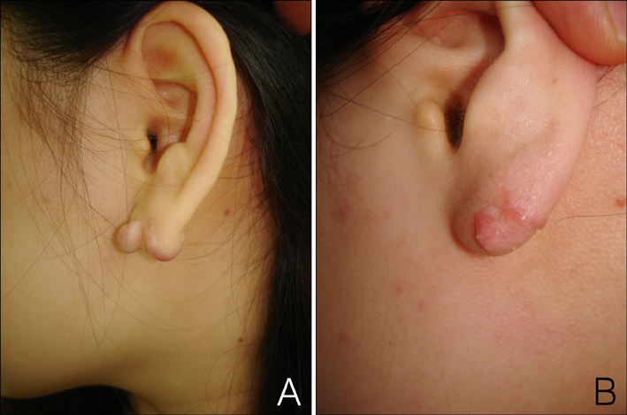
Fig. 2 (A) A lesion was observed on the left ear lobe. (B) Radiotherapy was performed 7 days after surgery. A small, recurrent, hypervascular lesion re-grew after radiotherapy. Steroid injection therapy was administered to the recurrent lesion.
Reference
-
1. Marneros AG, Krieg T. Keloids--clinical diagnosis, pathogenesis, and treatment options. J Dtsch Dermatol Ges. 2004; 2:905–913.2. Brissett AE, Sherris DA. Scar contractures, hypertrophic scars, and keloids. Facial Plast Surg. 2001; 17:263–272.
Article3. Lee JY, Yang CC, Chao SC, Wong TW. Histopathological differential diagnosis of keloid and hypertrophic scar. Am J Dermatopathol. 2004; 26:379–384.
Article4. Jung JY, Roh MR, Kwon YS, Chung KY. Surgery and perioperative intralesional corticosteroid injection for treating earlobe keloids: a korean experience. Ann Dermatol. 2009; 21:221–225.
Article5. Clark JA, Turner ML, Howard L, Stanescu H, Kleta R, Kopp JB. Description of familial keloids in five pedigrees: evidence for autosomal dominant inheritance and phenotypic heterogeneity. BMC Dermatol. 2009; 9:8.
Article6. Omo-Dare P. Genetic studies on keloid. J Natl Med Assoc. 1975; 67:428–432.7. Ogawa R, Mitsuhashi K, Hyakusoku H, Miyashita T. Postoperative electron-beam irradiation therapy for keloids and hypertrophic scars: retrospective study of 147 cases followed for more than 18 months. Plast Reconstr Surg. 2003; 111:547–553. discussion 554-555.
Article8. van de Kar AL, Kreulen M, van Zuijlen PP, Oldenburger F. The results of surgical excision and adjuvant irradiation for therapy-resistant keloids: a prospective clinical outcome study. Plast Reconstr Surg. 2007; 119:2248–2254.
Article9. Midwood KS, Williams LV, Schwarzbauer JE. Tissue repair and the dynamics of the extracellular matrix. Int J Biochem Cell Biol. 2004; 36:1031–1037.
Article10. Chang HY, Sneddon JB, Alizadeh AA, Sood R, West RB, Montgomery K, et al. Gene expression signature of fibroblast serum response predicts human cancer progression: similarities between tumors and wounds. PLoS Biol. 2004; 2:E7.
Article11. Santoro MM, Gaudino G. Cellular and molecular facets of keratinocyte reepithelization during wound healing. Exp Cell Res. 2005; 304:274–286.
Article12. Fujiwara M, Muragaki Y, Ooshima A. Keloid-derived fibroblasts show increased secretion of factors involved in collagen turnover and depend on matrix metalloproteinase for migration. Br J Dermatol. 2005; 153:295–300.
Article13. Topol BM, Lewis VL Jr, Benveniste K. The use of antihistamine to retard the growth of fibroblasts derived from human skin, scar, and keloid. Plast Reconstr Surg. 1981; 68:227–232.
Article14. Caccialanza M, Piccinno R, Schiera A. Postoperative radiotherapy of keloids: a twenty-year experience. Eur J Dermatol. 2002; 12:58–62.15. Bischof M, Krempien R, Debus J, Treiber M. Postoperative electron beam radiotherapy for keloids: objective findings and patient satisfaction in self-assessment. Int J Dermatol. 2007; 46:971–975.
Article16. Rosen EM, Goldberg ID, Myrick KV, Levenson S. Radiation survival properties of cultured vascular smooth muscle cells. Radiat Res. 1984; 100:182–191.
Article17. Kingston HM. Genetics assessment and pedigree analysis. In : Rimoin DL, Connor JM, editors. Emery and Rimoin's principles and practice of medical genetics. 5th ed. Philadelphia: Churchill Livingstone;2007. p. 518–535.18. Marneros AG, Norris JE, Olsen BR, Reichenberger E. Clinical genetics of familial keloids. Arch Dermatol. 2001; 137:1429–1434.
Article19. Satish L, Lyons-Weiler J, Hebda PA, Wells A. Gene expression patterns in isolated keloid fibroblasts. Wound Repair Regen. 2006; 14:463–470.
Article20. Lim CP, Phan TT, Lim IJ, Cao X. Stat3 contributes to keloid pathogenesis via promoting collagen production, cell proliferation and migration. Oncogene. 2006; 25:5416–5425.
Article21. Bayat A, Bock O, Mrowietz U, Ollier WE, Ferguson MW. Genetic susceptibility to keloiddisease and transforming growth factor beta 2 polymorphisms. Br J Plast Surg. 2002; 55:283–286.
Article22. Kovalic JJ, Perez CA. Radiation therapy following keloidectomy: a 20-year experience. Int J Radiat Oncol Biol Phys. 1989; 17:77–80.23. Borok TL, Bray M, Sinclair I, Plafker J, LaBirth L, Rollins C. Role of ionizing irradiation for 393 keloids. Int J Radiat Oncol Biol Phys. 1988; 15:865–870.
Article
- Full Text Links
-
- Actions
-
Cited
- CITED
-
- Close
- Share
-
- Similar articles
-
- Therapeutic results and safety of postoperative radiotherapy for keloid after repeated Cesarean section in immediate postpartum period
- A Study on Dose Distribution at the Junction of (60)Co gamma-Ray and Elecron Beam in Postoperative Radiotherapy of Breast Cancer
- A Comparative Study on Recurrence of Earlobe Keloids after Postoperative Adjuvant Therapy
- Three Cases of Surgical Excision with Radiation Therapy in Auricular Keloids
- Dose Characteristics for IORT Applicator of ML-15MDX Electron Beam
