Histopathological Findings Are Associated with the Clinical Types of Psoriasis but Not with the Corresponding Lesional Psoriasis Severity Index
- Affiliations
-
- 1Department of Dermatology, Seoul National University Bundang Hospital, Seoul National University College of Medicine, Seongnam, Korea. swyoun@snu.ac.kr
- KMID: 2264833
- DOI: http://doi.org/10.5021/ad.2015.27.1.26
Abstract
- BACKGROUND
The assessment of the severity of psoriasis is often subjective because of the lack of quantitative laboratory diagnostic tools. Histopathological examination is the most commonly performed procedure for psoriasis diagnosis; however, it is usually descriptive. Thus, there is currently no quantitative method of determining psoriasis severity. The clinical types of psoriasis are correlated with the severity of the disease, and a lesional severity index, such as the psoriasis severity index (PSI), could be used as a quantitative tool for assessing gross severity.
OBJECTIVE
To correlate the histopathological findings of psoriasis with the PSI.
METHODS
Psoriatic lesions in 98 patients were evaluated. The lesions were classified into the guttate, papular, small plaque, and large plaque types according to morphology, and were scored according to the PSI. Ten common histopathological features of psoriasis were evaluated for correlation with gross severity.
RESULTS
The clinical types of psoriasis showed significant correlations with the histopathological severity. However, the PSI score showed no correlation with histopathological severity.
CONCLUSION
In the future, subjective gross assessment should be modified by using objective measuring devices with detailed scales, in order to correlate the findings with the histological severity.
Keyword
MeSH Terms
Figure
-
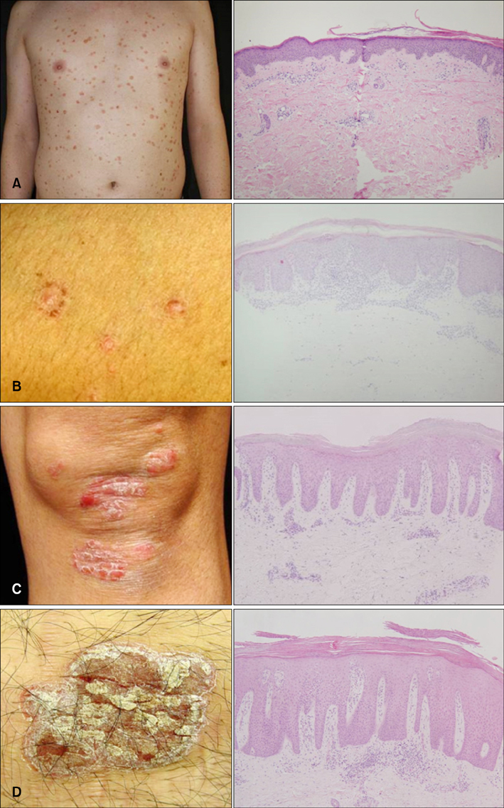
Fig. 1 Classification of the clinical types and histopathological features of the corresponding psoriatic lesions (H&E, ×100). (A) Guttate type, (B) papular type, (C) small plaque type, and (D) large plaque type.
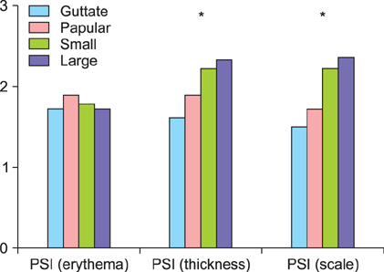
Fig. 2 Mean psoriasis severity index (PSI) scores in each clinical type of psoriasis, and comparisons according to the clinical types. Thickness and scale parameters showed statistically significant increases according to the gross morphological severity. Erythema showed no statistical significance. *p<0.05.

Fig. 3 Mean of histologic grades according to the clinical types of psoriasis. Most of the histologic findings showed significant change as the gross lesion size increased. *p<0.05.

Fig. 4 Psoriasis histopathologic score (PHS) according to the clinical types of psoriasis. Significant differences were found in different types of psoriasis (p<0.05).
Cited by 1 articles
-
Reduction of Inter-Rater and Intra-Rater Variability in Psoriasis Area and Severity Index Assessment by Photographic Training
Sang Woong Youn, Chong Won Choi, Bo Ri Kim, Je Byeong Chae
Ann Dermatol. 2015;27(5):557-562. doi: 10.5021/ad.2015.27.5.557.
Reference
-
1. Gudjonsson JE, Elder JT. Psoriasis. In : Goldsmith LA, Katz SI, Gilcrest BA, Paller AS, Leffell DJ, Wolff K, editors. Fitzpatrick's dermatology in general medicine. 8th ed. New York: McGraw-Hill;2012. p. 197–231.2. Mobini N, Toussant S, Kamino H. Noninfectious erythematous, papular, and squamous diseases. In : Elder DE, editor. Lever's histopathology of skin. 10th ed. Philadelphia: Lippincott Williams & Wilkins;2009. p. 169–203.3. Puzenat E, Bronsard V, Prey S, Gourraud PA, Aractingi S, Bagot M, et al. What are the best outcome measures for assessing plaque psoriasis severity? A systematic review of the literature. J Eur Acad Dermatol Venereol. 2010; 24:Suppl 2. 10–16.
Article4. Garduno J, Bhosle MJ, Balkrishnan R, Feldman SR. Measures used in specifying psoriasis lesion(s), global disease and quality of life: a systematic review. J Dermatolog Treat. 2007; 18:223–242.
Article5. Spuls PI, Lecluse LL, Poulsen ML, Bos JD, Stern RS, Nijsten T. How good are clinical severity and outcome measures for psoriasis?: quantitative evaluation in a systematic review. J Invest Dermatol. 2010; 130:933–943.
Article6. Kim JH, Kim BY, Choi JW, Kim SO, Lee HS, Park KC, et al. The objective evaluation of the severity of psoriatic scales with desquamation collecting tapes and image analysis. Skin Res Technol. 2012; 18:143–150.
Article7. Choi JW, Kwon SH, Youn JI, Youn SW. Objective measurements of erythema, elasticity and scale could overcome the inter- and intra-observer variations of subjective evaluations for psoriasis severity. Eur J Dermatol. 2013; 23:224–229.
Article8. Cox AJ, Watson W. Histological variations in lesions of psoriasis. Arch Dermatol. 1972; 106:503–506.
Article9. Griffin TD, Lattanand A, VanScott EJ. Clinical and histologic heterogeneity of psoriatic plaques. Therapeutic relevance. Arch Dermatol. 1988; 124:216–220.
Article10. Beek CH, van Reede EC. The nature and frequency of thehistological changes found in psoriasis vulgaris. Arch Dermatol Res. 1977; 257:255–264.
Article11. Trozak DJ. Histologic grading system for psoriasis vulgaris. Int J Dermatol. 1994; 33:380–381.
Article12. Ragaz A, Ackerman AB. Evolution, maturation, and regression of lesions of psoriasis. New observations and correlation of clinical and histologic findings. Am J Dermatopathol. 1979; 1:199–214.13. Murphy M, Kerr P, Grant-Kels JM. The histopathologic spectrum of psoriasis. Clin Dermatol. 2007; 25:524–528.
Article14. Ardigo M, Cota C, Berardesca E, González S. Concordance between in vivo reflectance confocal microscopy and histology in the evaluation of plaque psoriasis. J Eur Acad Dermatol Venereol. 2009; 23:660–667.
Article15. Morsy H, Kamp S, Thrane L, Behrendt N, Saunder B, Zayan H, et al. Optical coherence tomography imaging of psoriasis vulgaris: correlation with histology and disease severity. Arch Dermatol Res. 2010; 302:105–111.
Article16. Welzel J, Reinhardt C, Lankenau E, Winter C, Wolff HH. Changes in function and morphology of normal human skin: evaluation using optical coherence tomography. Br J Dermatol. 2004; 150:220–225.
Article17. Hoffmann K, Dirschka T, Schwarze H, el-Gammal S, Matthes U, Hoffmann A, et al. 20 MHz sonography, colorimetry and image analysis in the evaluation of psoriasis vulgaris. J Dermatol Sci. 1995; 9:103–110.
Article
- Full Text Links
-
- Actions
-
Cited
- CITED
-
- Close
- Share
-
- Similar articles
-
- An Inverse Relationship Between Ceramide Synthesis and Clinical Severity in Patients with Psoriasis
- Negative Correlation between CD163 Expression and Psoriasis Severity
- Relation between the Peripherofacial Psoriasis and Scalp Psoriasis
- Clinical Study on Psoriasis - 2 . Classification of Severity and Comparative Study by the Activity of Psoriasis
- Clinical Analysis of Nail Involvement in Psoriasis and Psoriatic Arthritis
