Korean J Gastroenterol.
2014 Jun;63(6):366-368. 10.4166/kjg.2014.63.6.366.
Pseudoepitheliomatous Hyperplasia Mimicking Esophageal Squamous Cell Carcinoma in a Patient with Lye-induced Esophageal Stricture
- Affiliations
-
- 1Department of Internal Medicine, Korea University College of Medicine, Seoul, Korea. leesw@kumc.or.kr
- KMID: 2234038
- DOI: http://doi.org/10.4166/kjg.2014.63.6.366
Abstract
- Pseudoepitheliomatous hyperplasia is a benign condition that may be caused by prolonged inflammation, chronic infection, and/or neoplastic conditions of the mucous membranes or skin. Due to its histological resemblance to well-differentiated squamous cell carcinoma, pseudoepitheliomatous hyperplasia may occasionally be misdiagnosed as squamous cell carcinoma. The importance of pseudoepitheliomatous hyperplasia is that it is a self-limited condition that must be distinguished from squamous cell carcinoma before invasive treatment. We report here on a rare case of esophageal pseudoepitheliomatous hyperplasia in a 67-year-old Korean woman with a lye-induced esophageal stricture. Although esophageal pseudoepitheliomatous hyperplasia is infrequently encountered, pseudoepitheliomatous hyperplasia should be considered in the differential diagnosis of esophageal lesions.
MeSH Terms
Figure
-
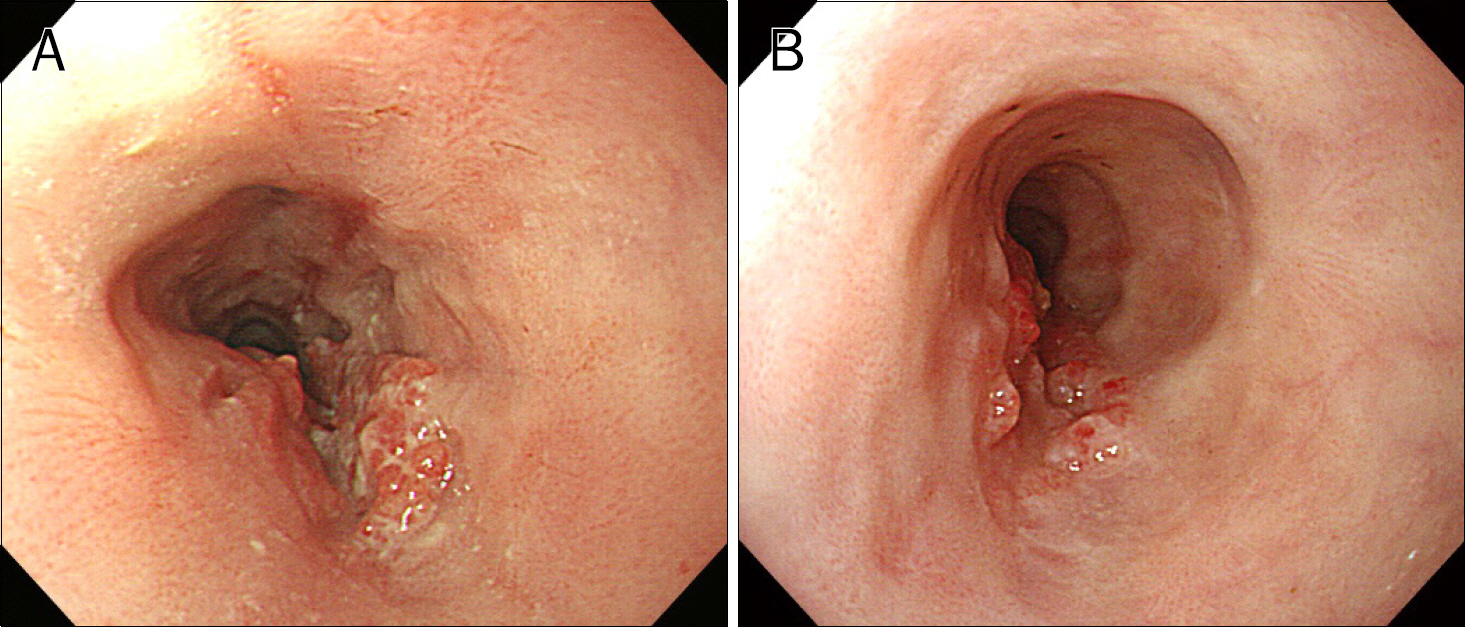
Fig. 1. Endoscopic view of the esophagus showing an intraluminal protruding mass 25 cm from the incisor teeth during: (A) an initial esophagogastroduodenoscopy; (B) a surveillance esophagogastroduodenoscopy performed after two months.
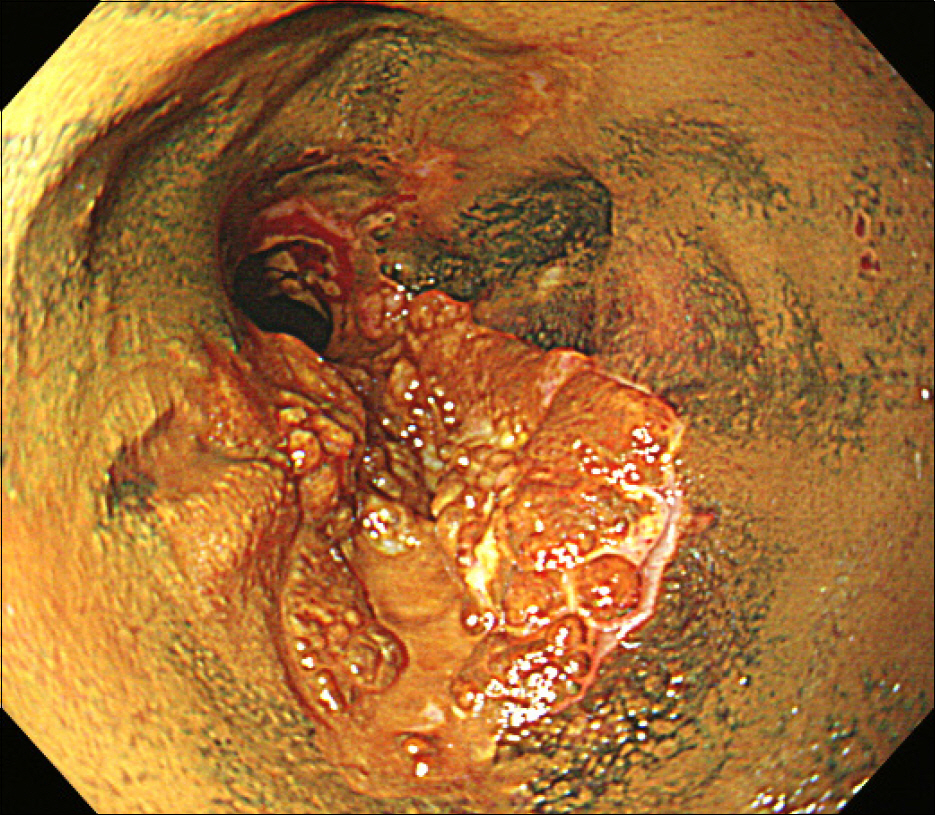
Fig. 2. Lugol chromoscopy showing the mucosal lesion stained with Lugol's solution.
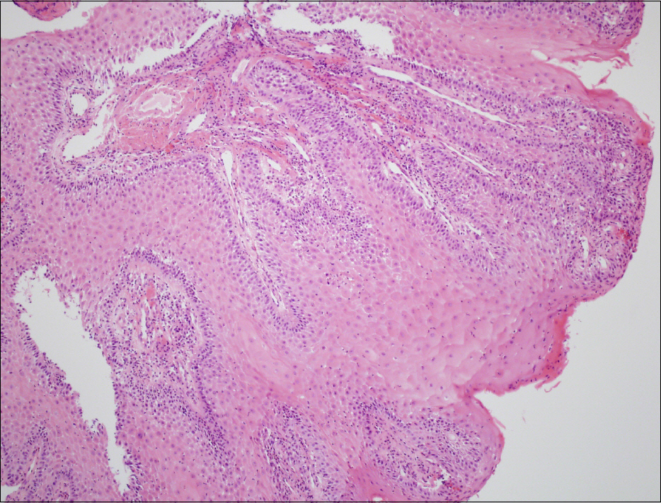
Fig. 3. Histological view of the esophageal mucosal lesion showing prominent hyperplasia of the epithelium (H&E, ×100).
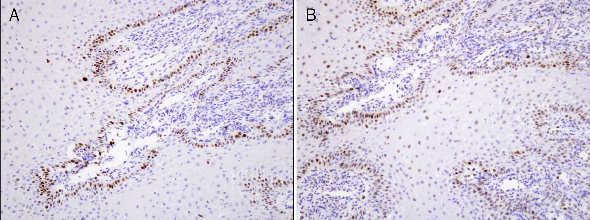
Fig. 4. Immunohistochemical view of the esophageal mucosal lesion showing staining limited to the basal layer of benign epithelium with: (A) Ki67 immunostain (×200); (B) p53 immunostain (×200).
Reference
-
References
1. Fu X, Jiang D, Chen W, Sun Bs T, Sheng Z. Pseudoepitheliomatous hyperplasia formation after skin injury. Wound Repair Regen. 2007; 15:39–46.
Article2. Zayour M, Lazova R. Pseudoepitheliomatous hyperplasia: a review. Am J Dermatopathol. 2011; 33:112–122.
Article3. Biswas A, Gey van Pittius D, Stephens M, Smith AG. Recurrent primary cutaneous lymphoma with florid pseudoepitheliomatous hyperplasia masquerading as squamous cell carcinoma. Histopathology. 2008; 52:755–758.
Article4. Lee YS, Teh M. p53 expression in pseudoepitheliomatous hyperplasia, keratoacanthoma, and squamous cell carcinoma of skin. Cancer. 1994; 73:2317–2323.
Article5. Zarovnaya E, Black C. Distinguishing pseudoepitheliomatous hyperplasia from squamous cell carcinoma in mucosal biopsy specimens from the head and neck. Arch Pathol Lab Med. 2005; 129:1032–1036.
Article
- Full Text Links
-
- Actions
-
Cited
- CITED
-
- Close
- Share
-
- Similar articles
-
- Cutaneous Metastasis of Esophageal Squamous Cell Carcinoma Mimicking Benign Soft Tissue Tumor
- Balloon catheter dilatation of esophageal strictures
- Esophageal foreign body extraction by biliary stone basket: a case report
- A Case of Esophageal Stricture by Lye that Treated with Esophageal Endoscopic Endoprosthesis
- A Case of Refractory Esophageal Stricture Induced by Lye Ingestion and Treated by Temporary Placement of Newly Designed Self-Expanding Metal Stent and Wetting with Mitomycin C
