Korean Circ J.
2010 Jul;40(7):354-355. 10.4070/kcj.2010.40.7.354.
External Mass Compressing the Left Atrium on Transthoracic Echocardiography
- Affiliations
-
- 1Division of Cardiology, Department of Internal Medicine, St. Vincent's Hospital, The Catholic University of Korea, Suwon, Korea. cmkim@cmcnu.or.kr
- KMID: 2225182
- DOI: http://doi.org/10.4070/kcj.2010.40.7.354
Abstract
- No abstract available.
MeSH Terms
Figure
-
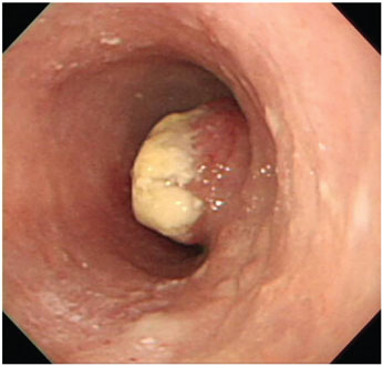
Fig. 1 Esophagogastroduodenoscopy shows a 3.0×2.4×8.0 cm esophageal mass.
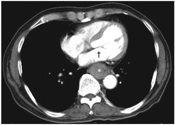
Fig. 2 Chest CT reveals an esophageal mass (*) which compresses the left atrium (†).
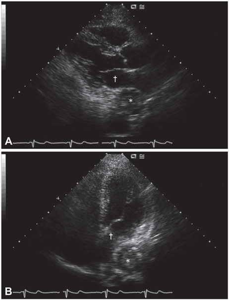
Fig. 3 Two-dimensional echocardiographic parasternal long axis view (A) and apical four-chamber view (B). A round and well-demarcated esophageal mass (*) compresses the left atrium (†), which resembles the image on chest CT.
Reference
-
1. Shah A, Tunick PA, Greaney E, Pfeffer RD, Kronzon I. Diagnosis of esophageal carcinoma because of findings on transesophageal echocardiography. J Am Soc Echocardiogr. 2001. 14:1134–1136.2. Im E, Shim CY, Hwang HJ, et al. Transthoracic echocardiographic detection, differential diagnosis, and follow-up of esophageal hematoma. Korean Circ J. 2007. 37:666–670.3. Walpot J, Amsel B, Pasteuning WH, Olree M. Left atrial compression by dissecting aneurysm of the ascending aorta. J Am Soc Echocardiogr. 2007. 20:1220.e4–1220.e6.4. Pehlivan Y, Sevinc A, Ozer O, Sari I, Davutoglu V. Mediastinal testicular tumor compressing the left atrium in a young male presenting initially with symptoms of left heart failure. Intern Med. 2009. 48:169–171.
- Full Text Links
-
- Actions
-
Cited
- CITED
-
- Close
- Share
-
- Similar articles
-
- Left Atrial Mass with Stalk: Thrombus or Myxoma?
- Right Atrial Blood Cyst Mimicking a Vegetative Mass
- Left Atrium Compressed by a Traumatic Focal Aneurysm of the Thoracic Aorta
- A Case of Esophageal Achalasia Compressing Left Atrium Diagnosed by Echocardiography in Patient with Acute Chest Pain
- Cerebellar Embolization in Patients with Heart Murmur
