Right Atrial Blood Cyst Mimicking a Vegetative Mass
- Affiliations
-
- 1Department of Cardiology, Pusan National University Hospital, Pusan National University School of Medicine, Busan, Korea
- KMID: 2517224
- DOI: http://doi.org/10.7180/kmj.2021.36.1.40
Abstract
- A 79-year-old woman presented to another hospital with complaints of right leg pain. Computed tomography and magnetic resonance imaging of the spine was performed in the other hospital, which showed an abscess in the right iliacus muscle. She was referred to our hospital because of a mass in the right atrium on echocardiography. Inflammatory markers were elevated, and Staphylococcus aureus were identified in blood cultures. Transthoracic echocardiography revealed a shaggy mass in the right atrium that resembled vegetation. Transesophageal echocardiography showed a large cystic mass with a hyperechoic lesion. After surgery, biopsy results indicated that it was a myxoid mass with cystic changes.
Keyword
Figure
-
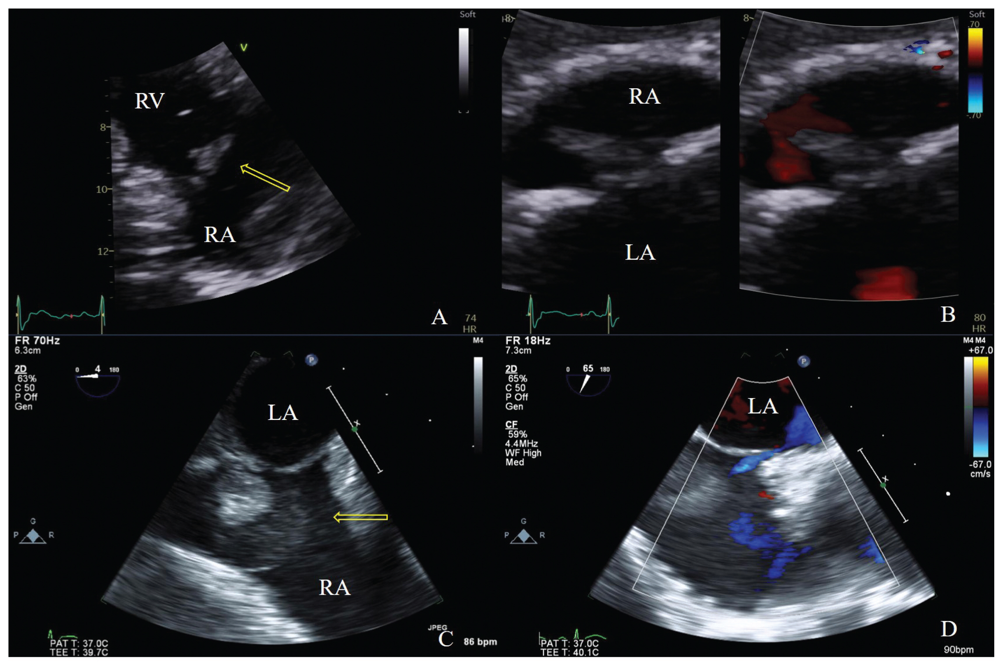
Fig. 1 (A) TTE in the parasternal RV inflow view showing a shaggy-appearing mass. (B) TTE in the subxiphoid view, showing a mass with color Doppler. (C) TEE shows a large hypoechoic mass and shaggy-appearing mass. (D) A patent foramen ovale is seen on TEE. TTE, transthoracic echocardiography; RV, right ventricular; TEE, transthoracic echocardiography

Fig. 2 Heart CT image showing a 2.5 cm mass arising from the interatrial septum of the RA. CT, computed tomography; RA, right atrium
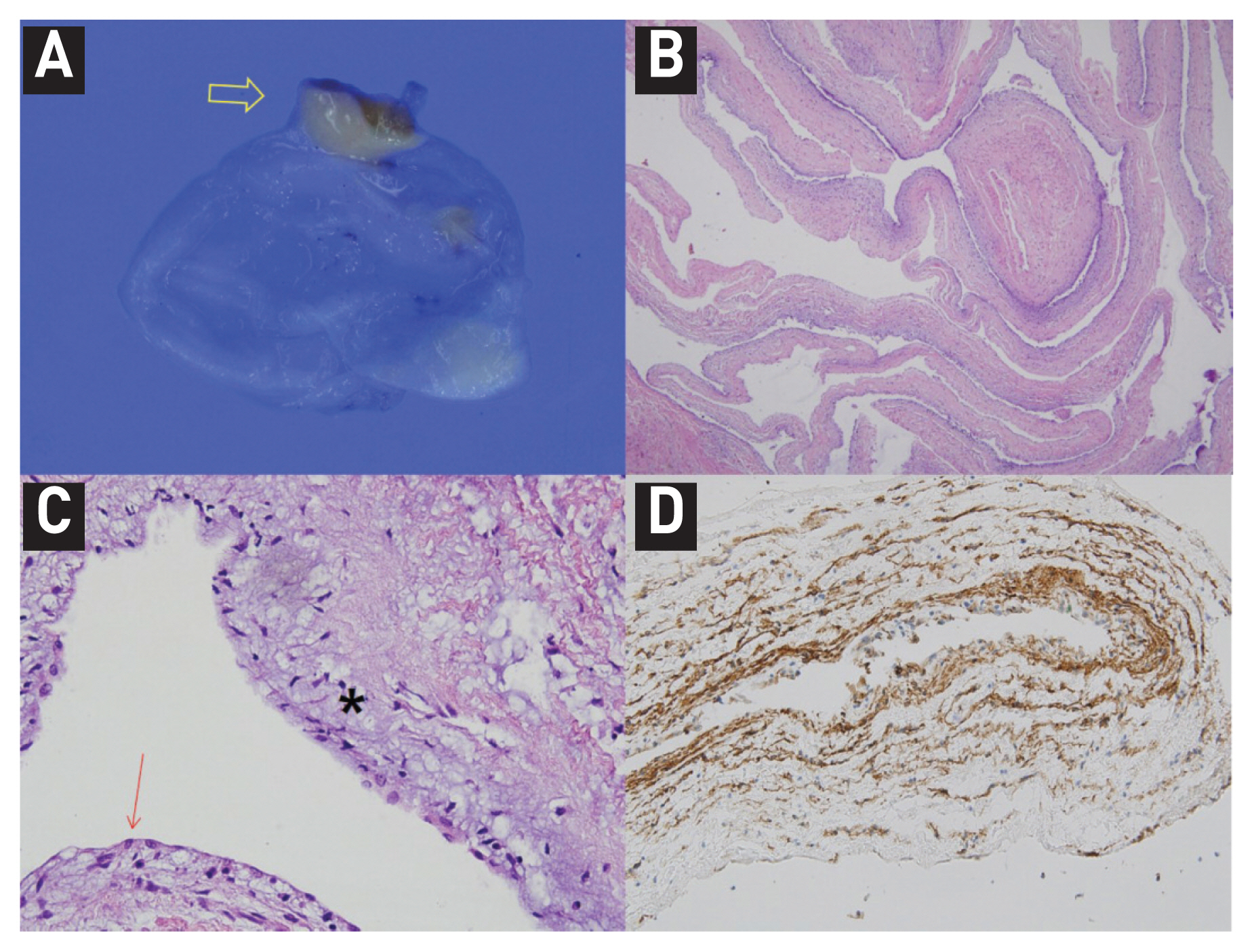
Fig. 3 (A) Gross findings of the resected RA mass. The cystic mass shows a translucent cystic wall without a solid nodule. The pedicle of the cystic mass is identified (arrow). (B) Microscopic examination of the resected specimen. A. At low magnification, the cystic wall shows myxoid changes in the surface aspect and is supported by a fibromuscular wall (H-E stained, x40). (C) At a high-power view (x400), the cystic wall appears composed of the flat surface epithelium and subepithelial myxoid stroma (flat surface epithelium; red arrow, myxoid stroma; star). (D) The surface epithelium and stellate or spindle-shaped stromal cells appear focally positive for CD34 which was often positive in myxoma10. RA, right atrium; H-E, hematoxylin and eosin
Reference
-
1. Roberts WC. Primary and secondary neoplasms of the heart. Am J Cardiol. 1997; 80:671–82.
Article2. Peters PJ, Reinhardt S. The echocardiographic evaluation of intracardiac masses: a review. J Am Soc Echocardiogr. 2006; 19:230–40.
Article3. Shakerian B, Jebelli M. Right Atrium Blood Cyst and Calcified Kernel in an Adult. Clin Med Insights Case Rep. 2019; 12:1–2.
Article4. Park MH, Jung SY, Youn HJ, Jin JY, Lee JH, Jung HO. Blood cyst of subvalvular apparatus of the mitral valve in an adult. J Cardiovasc Ultrasound. 2012; 20:146–9.
Article5. Lee WC, Huang MP, Fu M. Multiple intracardiac masses: myxoma, thrombus or metastasis: a case report. J Med Case Rep. 2015; 9:179.
Article6. Elsässer C. Bericht uber die ereignisse in der gebaranstalt des CatherinenHospital in Jahre 1844. Med Correspondenzblatt. 1844; 14:297.7. Otsuka H, Arinaga K, Fukuda T, Takaseya T, Shojima T, Takagi K, et al. Double Right Atrial Blood Cysts. Ann Thorac Surg. 2016; 101:e147–9.
Article8. Dumantepe M, Ak K, Mungan U, Alp I, Inan BK, Yilmaz AT. Blood cyst of the right ventricle presenting as recurrent high fever and chills in an adult. Ann Thorac Surg. 2009; 87:638–40.
Article9. Agac MT, Acar Z, Turan T, Karadag B, Kul S, Erkan H. Blood cyst of tricuspid valve: an incidental finding in a patient with ventricular septal defect. Eur J Echocardiogr. 2009; 10(5):88–9.
Article10. Wang JG, Li YJ, Liu H, Li NN, Zhao J, Xing XM. Clinicopathologic analysis of cardiac myxomas: Seven years’ experience with 61 patients. J Thorac Dis. 2012; 4:272–83.
- Full Text Links
-
- Actions
-
Cited
- CITED
-
- Close
- Share
-
- Similar articles
-
- Four Cases of Cystic Nature Adjacent to Right Atrial Wall by Two-Dimensional Echocardiography
- Life Expectancy of The Posttraumatic Persistent Vegetative State: Review of Literature and A Proposal
- Tibial Schwannoma Mimicking a Popliteal Cyst
- Epidermoid Cyst after Groin Flap Mimicking Malignancy
- Anterior stafne bone cyst mimicking periapical cyst: a case report
