A Case of In-Stent Neointimal Plaque Rupture 10 Years After Bare Metal Stent Implantation: Intravascular Ultrasound and Optical Coherence Tomographic Findings
- Affiliations
-
- 1Division of Cardiology, Department of Internal Medicine, Keimyung University Dongsan Medical Center, Daegu, Korea. shur@dsmc.or.kr
- KMID: 2225076
- DOI: http://doi.org/10.4070/kcj.2011.41.11.671
Abstract
- Neointimal hyperplasia mainly develops within several months of coronary stent deployment, after which it stabilizes. Although it was widely accepted, particularly during the bare-metal stent (BMS) era, that in-stent restenosis (ISR) generally does not present as an acute coronary syndrome (ACS), but rather as a gradual recurrence of angina symptoms, recent data have shown that a substantial number of patients with ISR present as ACS. There has also been consistent postmortem evidence of plaque rupture secondary to atherosclerotic change within the neointima of a BMS. We report here a case of ACS in which intravascular ultrasound and optical coherent tomographic assessments revealed neointimal atherosclerotic change and ruptured plaque 10 years after BMS deployment.
Keyword
MeSH Terms
Figure
-
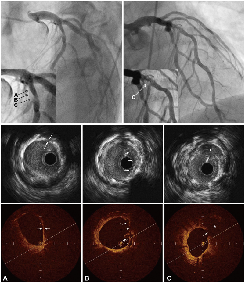
Fig. 1 Coronary angiogram (upper panel), intravascular ultrasound (middle panel) and optical coherence tomography (lower panel) images of the in-stent restenosis site in the proximal left anterior descending artery. A: in comparisons of IVUS and OCT images in the corresponding site, an atherosclerotic plaque extending from 12 to 5 o'clock contains regions consistent with fibrous tissue, and a homogenous signal-poor lesion possible representing lipid core with thin cap fibrous atheroma (TFCA) was clearly shown in OCT (arrow). Although this fibrous atheromatous lesion was also apparent in the IVUS image from the same site, it was difficult to identify the presence of TFCA and lipid core. The minimal cap thickness at the region measured 60 m by OCT. B: immediately proximal to the ruptured plaque, the OCT image showed multiple disrupted intimal flaps and a subtle ulcerated palque (arrow) that was not clearly seen in the corresponding IVUS image. C: an obvious intimal rupture (arrow) with large cavitary change (*) was also clearly identified by OCT compard with corresponding IVUS image.
Reference
-
1. Chen MS, John JM, Chew DP, Lee DS, Ellis SG, Bhatt DL. Bare metal stent restenosis is not a benign clinical entity. Am Heart J. 2006. 151:1260–1264.2. Doyle B, Rihal CS, O'Sullivan CJ, et al. Outcomes of stent thrombosis and restenosis during extended follow-up of patients treated with bare-metal coronary stents. Circulation. 2007. 116:2391–2398.3. Fineschi M, Carrera A, Gori T. Atheromatous degeneration of the neointima in a bare metal stent: intravascular ultrasound evidence. J Cardiovasc Med (Hagerstown). 2009. 10:572–573.4. Baek EK, Park SH, Kwon KH, Shim EJ, Jo JY. A case of in-stent plaque rupture presenting as an acute myocardial infarction. Korean Circ J. 2008. 38:432–435.5. Kume T, Akasaka T, Kawamoto T, et al. Assessment of coronary arterial plaque by optical coherence tomography. Am J Cardiol. 2006. 97:1172–1175.6. Jang IK, Bouma BE, Kang DH, et al. Visualization of coronary atherosclerotic plaques in patients using optical coherence tomography: comparison with intravascular ultrasound. J Am Coll Cardiol. 2002. 39:604–609.7. Komatsu R, Ueda M, Naruko T, Kojima A, Becker AE. Neointimal tissue response at sites of coronary stenting in humans: macroscopic, histological, and immunohistochemical analyses. Circulation. 1998. 98:224–233.8. Sangiorgi G, Taylor AJ, Farb A, et al. Histopathology of postpercutaneous transluminal coronary angioplasty remodeling in human coronary arteries. Am Heart J. 1999. 138:681–687.9. Chung IM, Gold HK, Schwartz SM, Ikari Y, Reidy MA, Wight TN. Enhanced extracellular matrix accumulation in restenosis of coronary arteries after stent deployment. J Am Coll Cardiol. 2002. 40:2072–2081.10. Lee CW, Kang SJ, Park DW, et al. Intravascular ultrasound findings in patients with very late stent thrombosis after either drug-eluting or bare-metal stent implantation. J Am Coll Cardiol. 2010. 55:1936–1942.11. Nakazawa G, Otsuka F, Nakano M, et al. The pathology of neoatherosclerosis in human coronary implants bare-metal and drug-eluting stents. J Am Coll Cardiol. 2011. 57:1314–1322.
- Full Text Links
-
- Actions
-
Cited
- CITED
-
- Close
- Share
-
- Similar articles
-
- Identification of Vulnerable Plaque in a Stented Coronary Segment 17 Years after Implantation Using Optical Coherence Tomography
- A Case of In-Stent Plaque Rupture Presenting as an Acute Myocardial Infarction
- A Newly Formed and Ruptured Atheromatous Plaque within Neointima after Drug-Eluting Stent Implantation: 2-Year Follow-Up Intravascular Ultrasound and Optical Coherence Tomography Studies
- Stent Evaluation with Optical Coherence Tomography
- Intravascular imaging analysis of a drug-eluting balloon followed by a bare metal stent compared to a drug-eluting stent for treatment of de novo lesions
