Korean Circ J.
2015 Nov;45(6):451-456. 10.4070/kcj.2015.45.6.451.
Radiation Exposure in Coronary Angiography: A Comparison of Cineangiography and Fluorography
- Affiliations
-
- 1Cardiovascular Center, Pusan National University Yangsan Hospital, Division of Cardiology, Department of Internal Medicine, Pusan National University School of Medicine, Yangsan, Korea. ptca82@gmail.com
- KMID: 2223782
- DOI: http://doi.org/10.4070/kcj.2015.45.6.451
Abstract
- BACKGROUND AND OBJECTIVES
Coronary angiography (CAG) is the gold standard for diagnosing coronary artery disease. However, exposure to ionizing radiation delivered during CAG has various negative biological effects on humans. In this study, there was an evaluation of whether fluorography resulted in decreased radiation exposure, as compared with cineangiography.
SUBJECTS AND METHODS
Fifty-five patients were prospectively enrolled and divided into two CAG groups, in accordance with the operator's professional discretion: a conventional cineangiography group versus a fluorography group. Fluorography refers to the photography of fluoroscopic images that are retrospectively stored, e.g., using the "Store fluoro" function of the Siemens cardiac angiography system. The primary outcomes included the air kinetic energy released per unit mass {air kerma (AK) mGy} and the dose (kerma)-area product (DAP; microGy . m2), both measured using built-in software in the Siemens system. The secondary outcomes included the total procedure time and amount of contrast agent used with each CAG method.
RESULTS
The total AK and DAP were significantly lower in the fluorography group (159.3+/-64.9 mGy and 1337.9+/-629.6 microGy . m2, respectively) than in the cineangiography group (326.9+/-107.5 mGy and 2341.1+/-849.9 microGy . m2, respectively; p=0.000 for both). The total procedure time (cineangiography vs. fluorography, 12.8+/-4.7 vs. 12.5+/-2.9 min; p=0.779) and contrast agent amount (136.1+/-28.3 vs. 126.3+/-25.7, p=0.214) were comparable between the two groups.
CONCLUSION
Fluorography is a useful method to decrease the radiation exposure in selected patients requiring CAG.
MeSH Terms
Figure
-
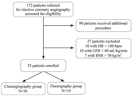
Fig. 1 Screening and enrollment of patients. After applying the exclusion criteria, a total of 55 patients were enrolled. HR: heart rate, GFR: glomerular filtration rate, BMI: body mass index.
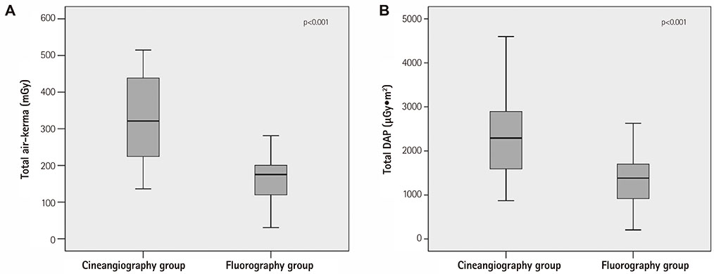
Fig. 2 Total air kerma and dose-area product values. (A) The total air kerma and (B) total dosearea product values were significantly lower in the fluorography group than in the cineangiography group. DAP: dose-area product.
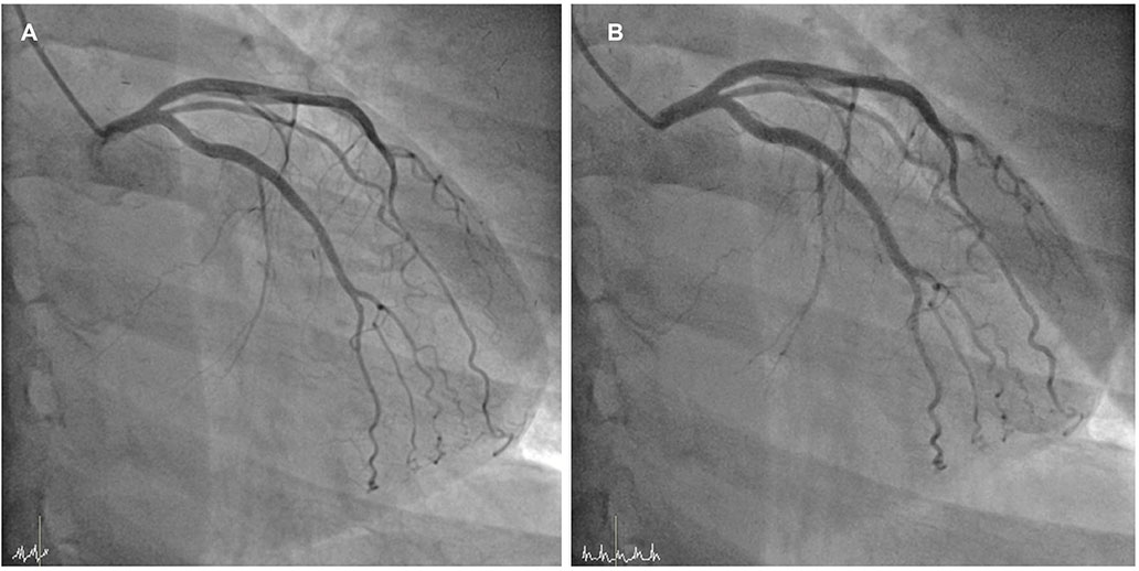
Fig. 3 Examples of angiographic images. These are angiographic images acquired through (A) cineangiography and (B) fluorography. The fluorography image (B) is comparable with the cineangiography image (A) and shows adequate details for decision-making.
Reference
-
1. Nickoloff EL, Lu ZF, Dutta A, So J, Balter S, Moses J. Influence of flat-panel fluoroscopic equipment variables on cardiac radiation doses. Cardiovasc Intervent Radiol. 2007; 30:169–176.2. Smith-Bindman R, Miglioretti DL, Johnson E, et al. Use of diagnostic imaging studies and associated radiation exposure for patients enrolled in large integrated health care systems, 1996-2010. JAMA. 2010; 307:2400–2409.3. Cousins C, Miller DL, Bernardi G, et al. ICRP PUBLICATION 120: radiological protection in cardiology. Ann ICRP. 2013; 42:1–125.4. Poll LW, Cohnen M, Brachten S, Ewen K, Mödder U. Dose reduction in multi-slice CT of the heart by use of ECG-controlled tube current modulation ("ECG pulsing"): phantom measurements. Rofo. 2002; 174:1500–1505.5. Delewi R, Hoebers LP, Råmunddal T, et al. Clinical and procedural characteristics associated with higher radiation exposure during percutaneous coronary interventions and coronary angiography. Circ Cardiovasc Interv. 2013; 6:501–506.6. Roguin A, Goldstein J, Bar O, Goldstein JA. Brain and neck tumors among physicians performing interventional procedures. Am J Cardiol. 2013; 111:1368–1372.7. Shah B, Mai X, Tummala L, et al. Effectiveness of fluorography versus cineangiography at reducing radiation exposure during diagnostic coronary angiography. Am J Cardiol. 2014; 113:1093–1098.8. Olcay A, Guler E, Karaca IO, et al. Comparison of fluoro and cine angiographic modes in coronary stenting procedure: a preliminary feasibility study. Int J Cardiol. 2014; 177:595–596.
- Full Text Links
-
- Actions
-
Cited
- CITED
-
- Close
- Share
-
- Similar articles
-
- Diagnostic value of cardiac cineangiography
- Coronary Artery Calcification Its Incidence and Significance in Patients Detected by Cineangiography
- Six Cases of Myocardial Infarction with Angiographically Normal or Near Normal Coronary Arteries
- Subacute Radiation Dermatitis due to Fluoroscopy during Cardiac Intervention
- The comparison of coronary arterial dimensions measured by cross-sextional echocardiography with values obtained by coronary angiography in Kawasaki disease
