Esophageal Cancer in Esophageal Diverticula Associated with Achalasia
- Affiliations
-
- 1Department of Internal Medicine, Gangnam Severance Hospital, Yonsei University College of Medicine, Seoul, Korea. HJPARK21@yuhs.ac
- 2Department of Thoracic Surgery, Gangnam Severance Hospital, Yonsei University College of Medicine, Seoul, Korea.
- 3Department of Diagnostic Pathology, Gangnam Severance Hospital, Yonsei University College of Medicine, Seoul, Korea.
- KMID: 2221762
- DOI: http://doi.org/10.5946/ce.2015.48.1.70
Abstract
- The simultaneous occurrence of achalasia and esophageal diverticula is rare. Here, we report the case of a 68-year-old man with multiple esophageal diverticula associated with achalasia who was later diagnosed with early esophageal cancer. He initially presented with dysphagia and dyspepsia, and injection of botulinum toxin to the lower esophageal sphincter relieved his symptoms. Five years later, however, the patient presented with worsening of symptoms, and esophagogastroduodenoscopy (EGD) was performed. The endoscopic findings showed multifocal lugol-voiding lesions identified as moderate dysplasia. We decided to use photodynamic therapy to treat the multifocal dysplastic lesions. At follow-up EGD 2 months after photodynamic therapy, more lugol-voiding lesions representing a squamous cell carcinoma in situ were found. The patient ultimately underwent surgery for the treatment of recurrent esophageal multifocal neoplasia. After a follow-up period of 3 years, the patient showed a good outcome without symptoms. To manage premalignant lesions such as achalasia with esophageal diverticula, clinicians should be cautious, but have an aggressive approach regarding endoscopic surveillance.
Keyword
MeSH Terms
Figure
-
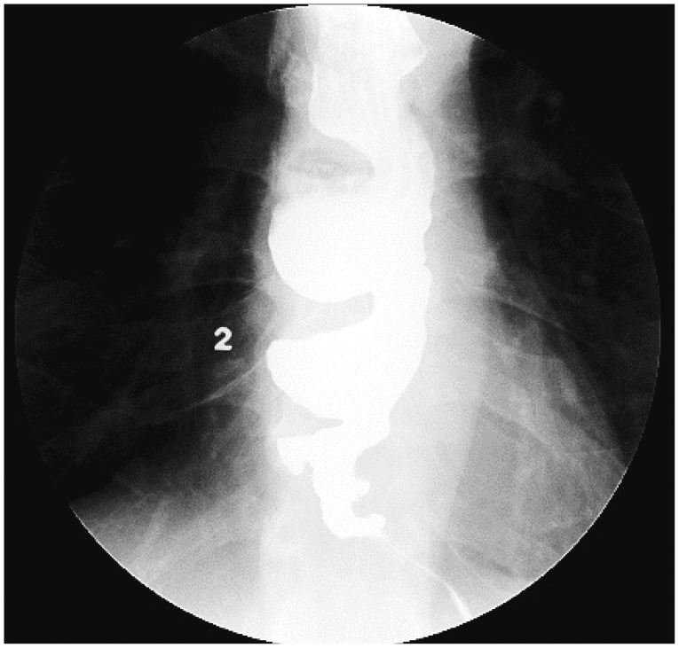
Fig. 1 Barium esophagography. View of a barium esophagogram showing multiple esophageal diverticula and a bird-beak appearance at the esophagogastric junction.
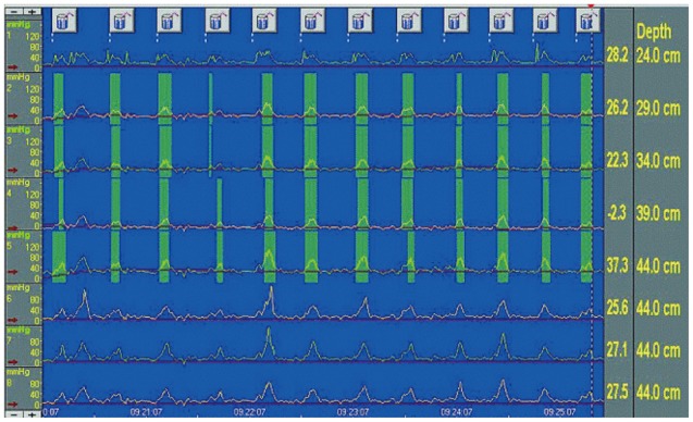
Fig. 2 Esophageal manometry. An esophageal manometric view shows the absence of peristalsis in the esophageal body and simultaneous contractions.
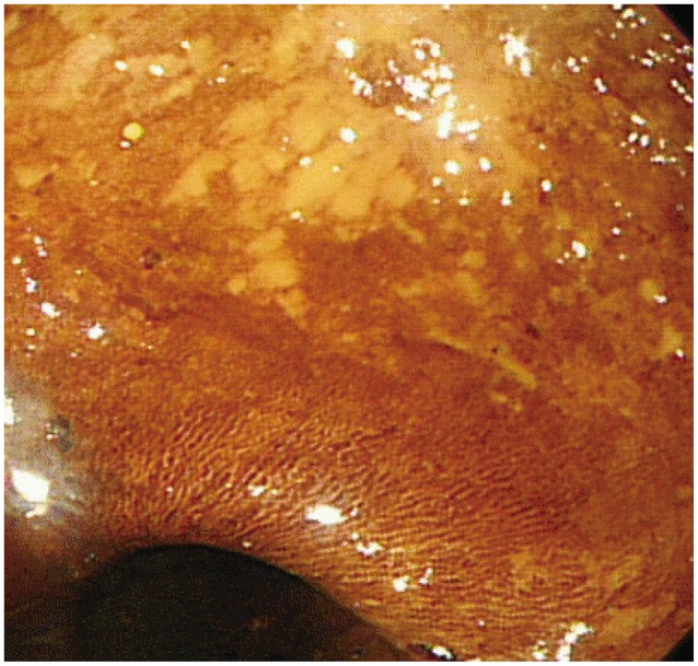
Fig. 3 Esophagogastroduodenography. Endoscopic view at 5 years after botulinum toxin injection therapy. A lugol-voiding lesion is seen at one of the multiple esophageal diverticula.
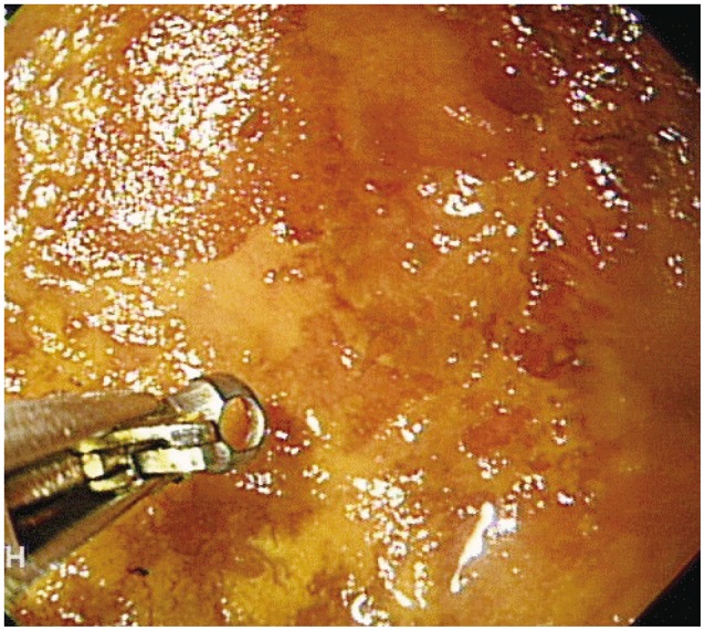
Fig. 4 Esophagogastroduodenoscopy after photodynamic therapy. Endoscopic view 2 months after photodynamic therapy. Another lugol-voiding lesion can be seen. Subsequently, a biopsy was performed.
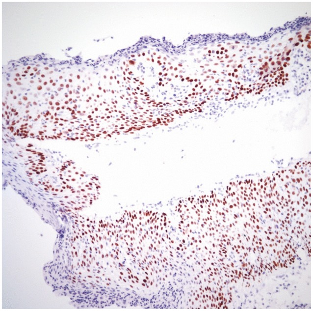
Fig. 5 Histologic findings. A biopsy specimen, from the lesion observed 2 months after photodynamic therapy, shown in Fig. 3 reveals a high-grade epithelial dysplasia and focal squamous cell carcinoma in situ (H&E stain, ×200).
Reference
-
1. Shin MS. Primary carcinoma arising in the epiphrenic esophageal diverticulum. South Med J. 1971; 64:1022–1024. PMID: 4998381.
Article2. Sen P, Kumar G, Bhattacharyya AK. Pharyngeal pouch: associations and complications. Eur Arch Otorhinolaryngol. 2006; 263:463–468. PMID: 16463064.
Article3. Honda H, Kume K, Tashiro M, et al. Early stage esophageal carcinoma in an epiphrenic diverticulum. Gastrointest Endosc. 2003; 57:980–982. PMID: 12776062.
Article4. Kimura H, Konishi K, Tsukioka Y, et al. Superficial esophageal carcinoma arising from the diverticulum of the esophagus. Endoscopy. 1997; 29:S53–S54. PMID: 9476779.
Article5. Kim Y, Kim JH, Kim C, Park H. Achalasia associated with multiple esophageal diverticula. Endoscopy. 2009; 41(Suppl 2):E47–E48. PMID: 19288420.
Article6. Goldenberg SP, Burrell M, Fette GG, Vos C, Traube M. Classic and vigorous achalasia: a comparison of manometric, radiographic, and clinical findings. Gastroenterology. 1991; 101:743–748. PMID: 1860637.
Article7. Dunaway PM, Wong RK. Risk and surveillance intervals for squamous cell carcinoma in achalasia. Gastrointest Endosc Clin N Am. 2001; 11:425–434. PMID: 11319071.
Article8. Leeuwenburgh I, Scholten P, Alderliesten J, et al. Long-term esophageal cancer risk in patients with primary achalasia: a prospective study. Am J Gastroenterol. 2010; 105:2144–2149. PMID: 20588263.
Article9. Loviscek LF, Cenoz MC, Badaloni AE, Agarinakazato O. Early cancer in achalasia. Dis Esophagus. 1998; 11:239–247. PMID: 10071806.
Article
- Full Text Links
-
- Actions
-
Cited
- CITED
-
- Close
- Share
-
- Similar articles
-
- A Case of Esophageal Carcinoma Following Esophagomyotomy for Achalasia
- Case of Concomitant Endoscopic Treatment of Achalasia with Superficial Esophageal Cancer
- A Case of Esophageal Carcinoma in a Patient with Primary Achalasia
- Esophageal Diverticulum
- Three Cases of the Secondary Achalasia induced by Malignant Tumor
