J Korean Ophthalmol Soc.
2013 Jun;54(6):958-961. 10.3341/jkos.2013.54.6.958.
A Case of a Congenital Lacrimal Outflow Dysgenesis with Supernumerary Lacrimal Puncta
- Affiliations
-
- 1Department of Ophthalmology, Chosun University College of Medicine, Gwangju, Korea. master@eyedoctors.co.kr
- KMID: 2217274
- DOI: http://doi.org/10.3341/jkos.2013.54.6.958
Abstract
- PURPOSE
To report a case of congenital lacrimal outflow dysgenesis with supernumerary lacrimal puncta.
CASE SUMMARY
A 45-years-old woman presented with chronic bilateral epiphora with no specific medical history. Slit-lamp examination revealed bilateral upper and lower double puncta. The accessory puncta were situated along the lid margin, medial to the normal one and had a typical slit configuration. Additionally, there was a lower canalicular system block that appeared unresponsive to simple probing. Bilateral endoscopic conjunctivodacryocystorhinostomy with Jones tube was performed. Chronic bilateral epiphora was relieved after bilateral endoscopic conjunctivodacryocystorhinostomy with Jones tube.
CONCLUSIONS
A case of congenital lacrimal outflow dysgenesis with supernumerary lacrimal puncta was observed, which has not been previously reported in Korea. Surgical repair of the congenital lacrimal outflow dysgenesis with supernumerary lacrimal puncta should be considered to achieve a functional recovery of the lacrimal drainage system.
Figure
-

Figure 1. Pre-operative photographs of canaliculi obstruction and lacrimal dysgenesis. Lacrimal probe was exposed through canaliculi. (A) Right upper puncta, (B) Left upper puncta, (C) Right lower puncta, (D) Left lower puncta.
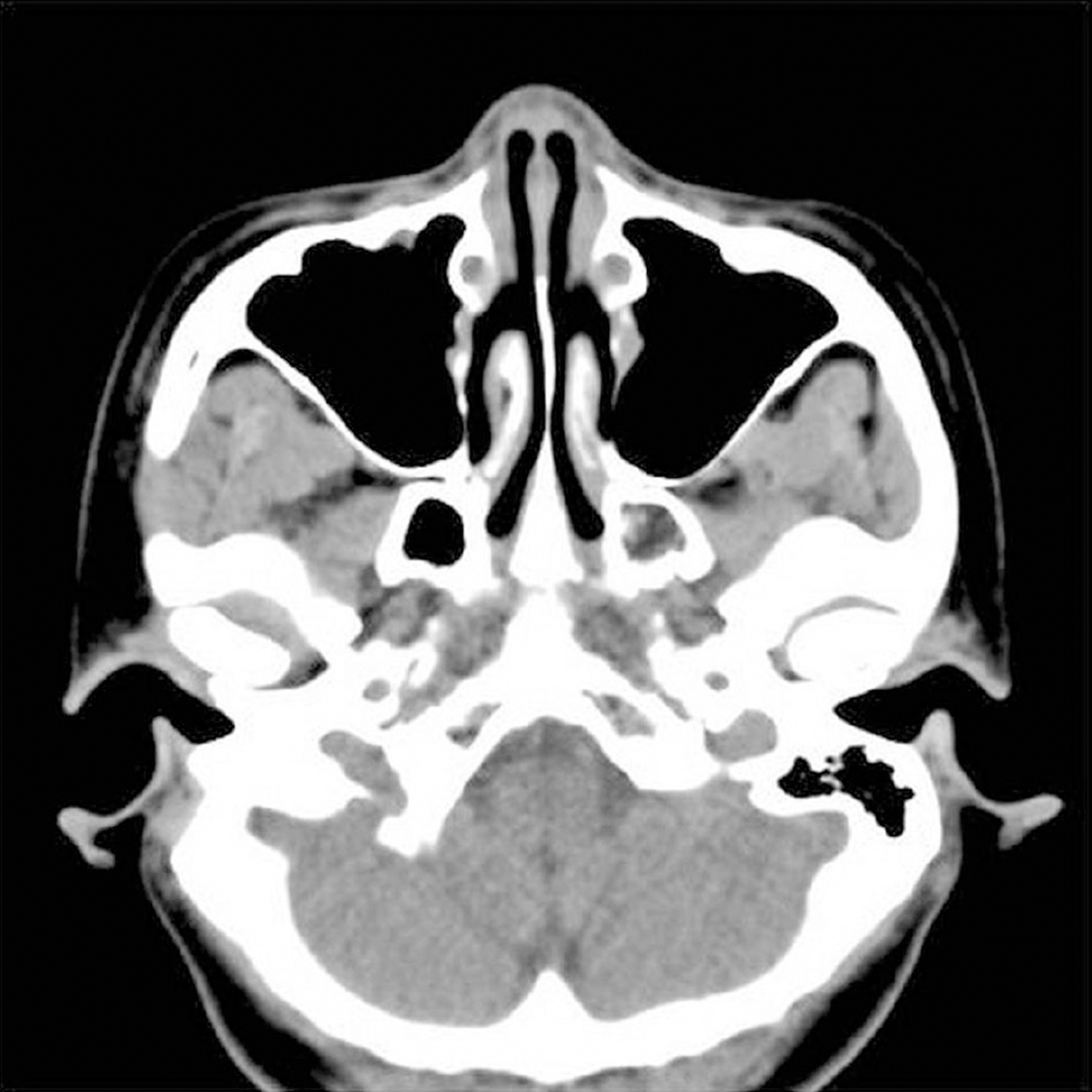
Figure 2. An axial computed tomography image shows bi-lateral nasolacrimal canal opacification.

Figure 3. Post-operative photographs of endoscopic conjunctivodacryocystorhinostomy state. The Lester-Jones’ tubes were inserted bilaterally. (A) Right eye, (B) Left eye.
Reference
-
References
1. Mackenzie W. A practical treatise on the diseases of the eye. 4th ed. London, England: Carter, Hendee;1854.2. Wicherkiewicz B. Congenital anomalies of the upper tear passages. Post Okul Krakow. 1905; March-April:596.3. Kleczkowski . Postep. Okulistyczny. 1908; 11.4. Bacskulin J. Double, triple and quadruple lacrimal puncta. Klin Monatsbl Augenheilkd. 1964; 144:418–28.5. Schoute GJ. Canalicule lacrymal surnumeraire. Arch Ophtalmol (Paris). 1901; 21:320–3.6. Kirk RC. Developmental anomalies of the lacrimal passages; a re-view of the literature and presentation of three unusual cases. Am J Ophthalmolol. 1956; 42:227–32.7. Carvill M. Congenital fistulae of the lacrimal canaliculi. Arch Ophthalmol. 1909; 38:585–90.8. Tooke FT. On so-called doubling of the puncta lacrimalia. Ophthal- mology: Essays, Abstracts and Reviews. 1910; 6:391–4.9. Chance B. Supernumerary punctum. Am J Ophthalmol. 1922; 5:297.10. Bothman L. Double puncta and double canaliculi of the upper lid. Am J Ophthalmol. 1932; 15:214–5.
Article11. Flom L, Levitt JM. Double lacrimal puncta and dacryops. AMA Arch Ophthalmol. 1955; 54:760–1.
Article12. Veirs ER. Disorders of the canaliculus. In: The Lacrimal System: Clinical Application. New York: Grune & Stratton;1955; 41–2.13. Mann I. The lids, lacrimal apparatus and orbital contents. In: Developmental Abnormalities of the Eye. 2nd ed.Philadelphia, PA: Lippincott;1957; 389–90.14. Duke-Elder S, Cook CA. Normal and abnormal development. Congenital deformities. In: Duke-Elder S, ed. System of Ophthal- mology. v. 3:part 2. London: Kimpton;1964; 928–34.15. Chignell AH. Double punctum and canaliculus. Am J Ophthalmol. 1968; 65:736–9.
Article16. Solomon A, Feiler-Ofry V, Lazar M. Congenital reduplication of the lacrimal punctum and canaliculus. Ann Ophthalmol. 1981; 13:727.17. Coats DK, McCreery KM, Plager DA. . Nasolacrimal outflow drainage anomalies in Down’s syndrome. Ophthalmology. 2003; 110:1437–41.
Article18. Yuen SJ, Oley C, Sullivan TJ. Lacrimal outflow dysgenesis. Op- hthalmology. 2004; 111:1782–90.
Article19. Bair PJ, Tsai YY, Lin JM. Congenital reduplication of the lacrimal punctum and canaliculus in a patient with dry eye. Ophthalmic Surg Lasers Imaging. 2004; 35:156–8.
Article20. Caccamise WC, Townes PL. Congenital absence of the lacrimal puncta associated with alacrima and aptyalism. Am J Ophthalmol. 1980; 89:62–5.
Article21. Town AE. Congenital absence of lacrimal puncta in three members of a family. Arch ophthalmol. 1943; 29:767–71.
Article22. Klapper SR, Jordan DR. Jones tube insertion in children with ca-nalicular agenesis. Ophthalmic Surg Lasers. 1999; 30:495–8.
Article23. Clauser L, Galiè M, Hassanipour A, Calabrese O. Saethre-Chotzen syndrome: review of the literature and report of a case. J Craniofac Surg. 2000; 11:480–6.
Article24. Miller MT, Deutsch TA, Cronin C, Keys CL. Amniotic bands as a cause of ocular anomalies. Am J Ophthalmol. 1987; 104:270–9.
Article25. Guion-Almeida ML, Rodini ES, Pereira SC, Richieri-Costa A. Amniotic bands and the EEC syndrome. Birth Defects Orig Artic Ser. 1996; 30:171–7.26. Hollsten DA, Katowitz JA. The ophthalmic manifestations and treatment of the amniotic band syndrome. Ophthal Plast Reconstr Surg. 1990; 6:1–15.27. McNab AA, Potts MJ, Welham RA. The EEC syndrome and its ocular manifestations. Br J Ophthalmol. 1989; 73:261–4.
Article28. Hay RJ, Wells RS. The syndrome of ankyloblepharon, ectodermal defects and cleft lip and palate: an autosomal dominant condition. Br J Dermatol. 1976; 94:277–89.
Article29. Jaffe MJ, Currie J, Schwankhaus JD, Sherins RJ. Ophthalmic midline dysgenesis in Kallmann syndrome. Ophthalmic Paediatr Genet. 1987; 8:171–4.
Article30. Markowitz GD, Handler LF, Katowitz JA. Congenital euryblepharon and nasolacrimal anomalies in a patient with Down syndrome. J Pediatr Ophthalmol Strabismus. 1994; 31:330–1.
Article31. Bowling BS, Chandna A. Superior lacrimal canalicular atresia and nasolacrimal duct obstruction in the CHARGE association. J Pediatr Ophthalmol Strabismus. 1994; 31:336–7.
Article32. Satchi K, McNab AA. Double lacrimal punta: Clinical presentation and potential mechanisms of epiphora. Ophthalmology. 2010; 117:180–3.e2.
