J Korean Ophthalmol Soc.
2019 Feb;60(2):187-189. 10.3341/jkos.2019.60.2.187.
An Unusual Case of Double Lacrimal Puncta
- Affiliations
-
- 1Department of Ophthalmology, Hallym University Kangdong Sacred Heart Hospital, Seoul, Korea. ophdrchoi@gmail.com
- KMID: 2438079
- DOI: http://doi.org/10.3341/jkos.2019.60.2.187
Abstract
- PURPOSE
Congenital double puncta are usually unilateral, and the accessory punctum exists on the medial side in a slit configuration that is distinct from the shape of the normal punctum. We report a case of an unusual case of double lacrimal puncta which the lateral, rather than the medial, punctum was judged to be the accessory punctum.
CASE SUMMARY
A 39-year-old male patient with no underlying disease and no ophthalmologic history visited our clinic with right eye epiphora of 2 weeks duration. On slit lamp examination, double puncta were observed in the right lower eyelid and the remaining puncta were normal. On lacrimal syringing test and dacryocystography were performed and revealed incomplete obstruction with partial narrowing of the nasolacrimal duct. Silicone tube intubation was performed through the right lower medial punctum and symptoms improved postoperatively.
CONCLUSIONS
The present case is an unusual case of double lacrimal puncta which has not been reported in Korea. Unlike the previous literature, the lateral, rather than the medial, punctum was judged to be the accessory punctum. Because accessory punctm can be present on the lateral side, it is necessary to distinguish between the accessory punctm and the main punctum through the accurate dacryocystography and lacrimal syringing test for the treatment of the patient.
MeSH Terms
Figure
-
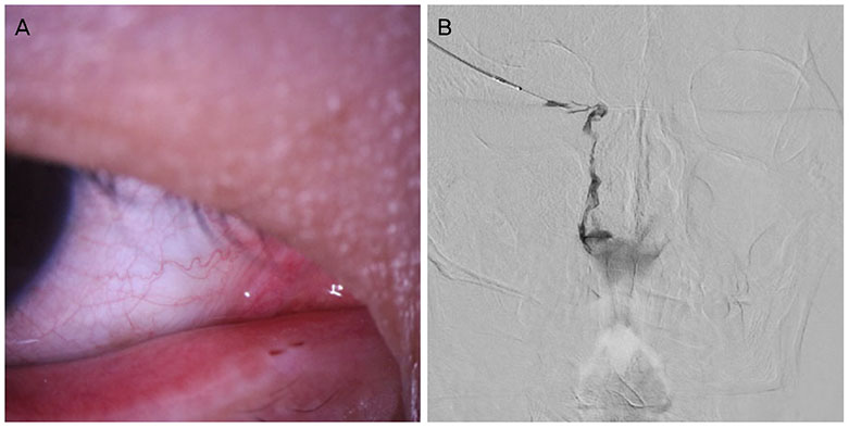
Figure 1 Slit lamp photograph and dacryocystography. (A) Slit lamp photograph of double puncta in the right lower eyelid. It is difficult to distinguish between main and accessory punctum on slit lamp examination because of their similar appearance. (B) Dacryocystography was performed via the right lower medial punctum, and revealed partial narrowing of the nasolacrimal duct.
Reference
-
1. Mackenzie W. A practical treatise on the disease of the eye. 4th ed. London: Blanchard & Lea;1854.2. Satchi K, McNab AA. Double lacrimal puncta: clinical presentation and potential mechanisms of epiphora. Ophthalmology. 2010; 117:180–183.
Article3. Lacroix Z, Bitton E. Supernumerary punctum: an unusual case of seeing double. Clin Exp Optom. 2015; 98:375–378.
Article4. Sevel D. Development and congenital abnormalities of the nasolacrimal apparatus. J Pediatr Ophthalmol Strabismus. 1981; 18:13–19.
Article5. Solomon A, Feiler-Ofry V, Lazar M. Congenital reduplication of the lacrimal punctum and canaliculus. Ann Ophthalmol. 1981; 13:727.
- Full Text Links
-
- Actions
-
Cited
- CITED
-
- Close
- Share
-
- Similar articles
-
- A Case of a Congenital Lacrimal Outflow Dysgenesis with Supernumerary Lacrimal Puncta
- Congenital Anomalies of Lacrimal Puncta
- A Case of Bilateral Dacryocystocele with Absence of the Lacrimal Puncta
- Tear Stasis Caused by Severely Protruded Lacrimal Puncta Treated by Novel Punctal Fixation Technique
- The Complex Choristoma with Ectopic Lacrimal Gland in Bilateral Bulbar Conjuntivas
