A Case of Retained Descemet's Membrane after Penetrating Keratoplasty
- Affiliations
-
- 1Department of Ophthalmology, Kangnam Sacred Heart Hospital, Hallym University College of Medicine, Seoul, Korea. schinn@hanmail.net
- KMID: 2217052
- DOI: http://doi.org/10.3341/jkos.2013.54.5.813
Abstract
- PURPOSE
To report a case of retained Descemet's membrane after penetrating keratoplasty (PKP).
CASE SUMMARY
A 64-year-old man visited our clinic, complaining of visual disturbance and corneal opacity in his right eye 40 years in duration. On the first visit, his best corrected visual acuity was hand movement on the right eye, and he underwent an uneventful PKP. On the postoperative first day, the patient's visual acuity was 20/200 and slit lamp examination showed a retained Descemet's membrane and pseudo-chamber behind the corneal graft. The corneal graft was edematous, but no intraocular inflammation was observed. The retained Descemet's membrane was surgically removed a quarter at a time. Sutures in one quadrant were removed; the retained Descemet's membrane was lifted with forceps, removed with scissors and knife, and then sutured again. Two months after PKP, the corneal graft remained clear and no intraocular inflammation was observed. An extracapsular cataract extraction (ECCE) was then successfully performed with posterior chamber lens implantation for the senile cataract in his right eye. After the 1-year follow-up, the status of the corneal graft remained clear with a single anterior chamber and best corrected visual acuity improved to 20/100.
CONCLUSIONS
Careful post-operative slit-lamp examination is considered important for diagnosis of retained Descemet's membrane after undergoing PKP, and surgical removal can be helpful for maintaining the corneal graft clear.
MeSH Terms
Figure
-
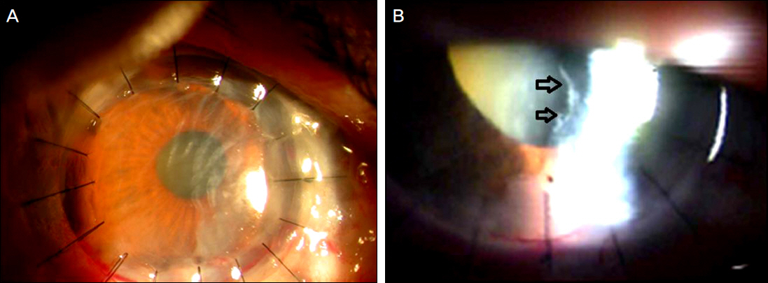
Figure 1. (A, B) One day after penetrating keratoplasty, slit-lamp examination shows a thin, fibrous membrane (arrows) behind the graft.

Figure 2. Representative OCT imaging of case 1. In the first postoperative day, OCT reveals retro-corneal membrane (arrows) as retained Descemet’s membrane maintaining its continuity with recipient cornea and also showing breach in the membrane. OCT = optical coherence tomography.
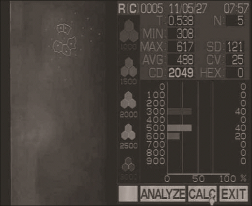
Figure 3. Endothelial cell count was 2049 cells/mm2 at the postoperative 2 months.
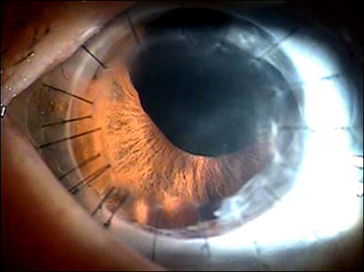
Figure 4. One year after penetrating keratoplasty, cornea graft has been clear.
Cited by 1 articles
-
Clinical Outcomes of Nd-YAG Laser Membranotomy in Retained Host Corneal Membrane after Keratoplasty
Yong Chan Kim, Jae Hyung Hwang, Man Soo Kim
J Korean Ophthalmol Soc. 2015;56(5):664-671. doi: 10.3341/jkos.2015.56.5.664.
Reference
-
References
1. Choi SH, Lee YW, Kim HM, et al. Epidemiologic studies of kera-toplasty in Korea. J Korean Ophthalmol Soc. 2006; 47:538–47.2. Jang JY, Woo JM, Yoon KC. Long-term results after three or more penetrating keratoplasties and risk factors for graft failure. J Korean Ophthalmol Soc. 2011; 52:1399–404.
Article3. Al-Qahtani FA. Scleral fixation of intraocular lenses combined with penetrating keratoplasty. J Cataract Refract Surg. 2010; 36:373–6.
Article4. Groh MJ, Seitz B, Händel A, Naumann GO. [Expulsive hemor-rhage in perforating keratoplasty--incidence and risk factors]. Klin Monbl Augenheilkd. 1999; 215:152–7.5. Chen YP, Lai PC, Chen PY, et al. Retained Descemet's membrane after penetrating keratoplasty. J Cataract Refract Surg. 2003; 29:1842–4.
Article6. Vengayil S, Vanathi M, Panda A, Khokhar S. Anterior segment OCT-based diagnosis and management of retained Descemet's membrane following penetrating keratoplasty. Cont Lens Anterior Eye. 2008; 31:161–3.
Article7. Thyagarajan S, Mearza AA, Falcon MG. Inadvertent retention of Descemet Membrane in penetrating keratoplasty. Cornea. 2006; 25:748–9.
Article8. Masket S, Tennen DG. Neodymium:YAG laser optical opening for retained Descemet's membrane after penetrating keratoplasty. J Cataract Refract Surg. 1996; 22:139–41.
Article9. Ide T, Yoo SH, Kymionis GD, et al. Double Descemet's membranes after penetrating keratoplasty with anterior segment optical coher-ence tomography. Ophthalmic Surg Lasers Imaging. 2008; 39:422–5.
Article10. Choi JS, Oh JY, Wee WR. A case of corneal endothelial deterio-ration associated with retained Descemet's membrane after pene-trating keratoplasty. Jpn J Ophthalmol. 2009; 53:653–5.
Article11. Sinha R, Vajpayee RB, Sharma N, et al. Trypan blue assisted desce-metorhexis for inadvertently retained Descemet's membranes after penetrating keratoplasty. Br J Ophthalmol. 2003; 87:654–5.
Article12. Kothari S, Kothari K, Parikh RS. Role of anterior segment optical coherence tomogram in Descemet's membrane detachment. Indian J Ophthalmol. 2011; 59:303–5.
Article13. Sonmez K, Ozcan PY, Altintas AG. Surgical repair of scrolled de-scemet’s membrane detachment with intracameral injection of 1.8% sodium hyaluronate. Int Ophthalmol. 2011; 31:421–3.
Article14. Patel SV, Hodge DO, Bourne WM. Corneal endothelium and post-operative outcomes 15 years after penetrating keratoplasty. Am J Ophthalmol. 2005; 139:311–9.
Article15. Arenas Archila E, Ramirez Cabrera MF, Mieth Alviar A. Double Descemet's membrane in penetrating keratoplasty. Refract Corneal Surg. 1993; 9:65–6.16. Steinemann TL, Henry K, Brown MF. Nd:YAG laser treatment of retained Descemet's membrane after penetrating keratoplasty. Ophthalmic Surg. 1995; 26:80–1.17. Henderson JW, Wolter JR. Separation of Descemet's membrane in keratoplasty. Am J Ophthalmol. 1968; 65:375–8.
Article
- Full Text Links
-
- Actions
-
Cited
- CITED
-
- Close
- Share
-
- Similar articles
-
- A Case of Double Descemet's Membrane after Penetrating Keratoplasty Converted from Deep Anterior Lamellar Keratoplasty
- Descemet Membrane Endothelial Keratoplasty after Penetrating Keratoplasty Graft Failure
- A Case of Herpes Simplex Keratitis after Descemet Membrane Endothelial Keratoplasty
- Early Result of Femtosecond Laser Assisted Descemet's Membrane Stripping Endothelial Keratoplasty
- A Case of Posterior Polymorphous Dystrophy
