Change in Central Macular Thickness after 2.2-mm Microincision Coaxial versus 2.75-mm Small Incision Cataract Surgery
- Affiliations
-
- 1Department of Ophthalmology, Bucheon St. Mary's Hospital, The Catholic University of Korea College of Medicine, Bucheon, Korea. eunchol@hanmail.net
- KMID: 2216863
- DOI: http://doi.org/10.3341/jkos.2014.55.10.1460
Abstract
- PURPOSE
To evaluate the central macular thickness and volume changes after conventional 2.75-mm small incision cataract surgery (SICS) and 2.2-mm microincision coaxial cataract surgery (MICS).
METHODS
We performed a retrospective chart review of 48 eyes undergoing uneventful phacoemulsification and divided the subjects into two groups, SICS and MICS. To evaluate the central macular thickness and volume changes after cataract surgery, optical coherence tomography (OCT) was used before and at one day, one week, one month, and two months postoperatively.
RESULTS
No statistically significant difference was found in the intraoperative phacoemulsification power, phaco time, or volume of intraoperative irrigation solution between the two groups (p > 0.05). The average central macular thickness increased in the MICS group compared to the SICS group at every postoperative time point, and the difference was statistically significant at postoperative one month (p = 0.04). The average central macular volume was elevated in the MICS group; however, the difference was not significant (p > 0.05).
CONCLUSIONS
Central macular thickness and volume change were greater in the MICS group compared to the SICS group, and the difference in central macular thickness between the two groups was significant at postoperative one month. Careful follow-up examinations should be performed using OCT at this postoperative time point, especially in patients who received cataract surgery with smaller incision size.
Keyword
MeSH Terms
Figure
-
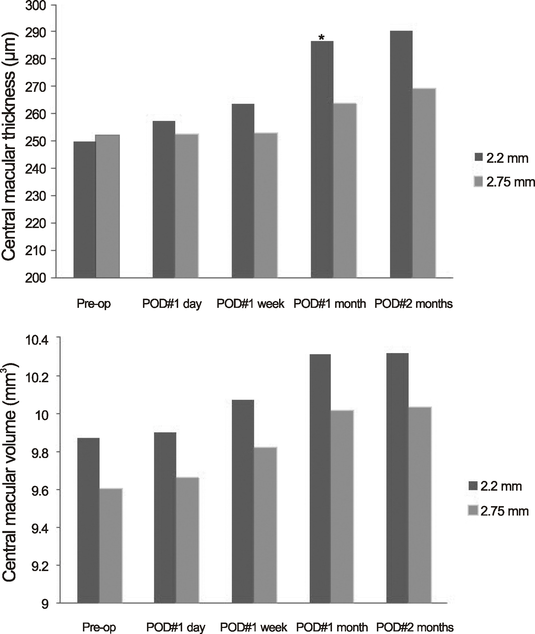
Figure 1. Central macular thickness and central macular volume change at each postoperative time period (student t-test p < 0.05*). POD = postoperative date.
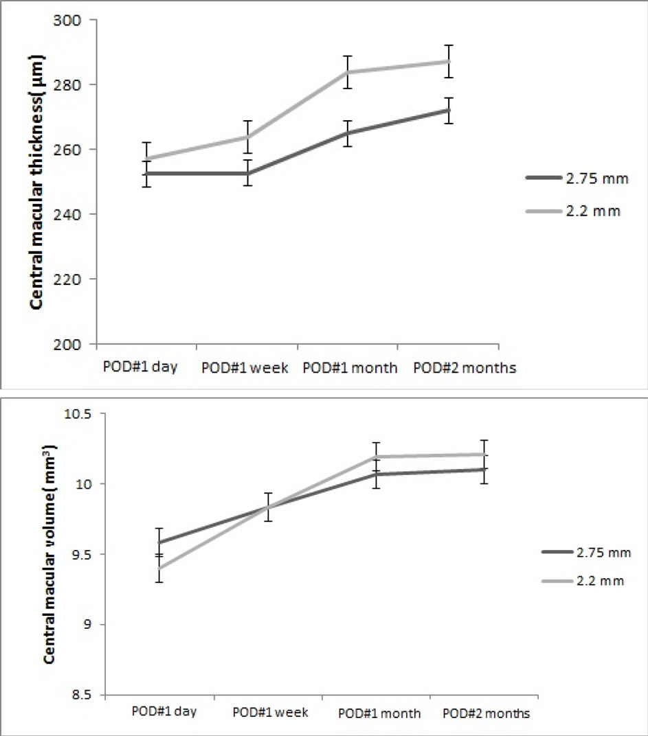
Figure 2. Analysis of central macular thickness and central macular volume change in time course at each postoperative period (repeated measures ANOVA; Central macular thickness; Small incision cataract surgery (SICS) group p = 0.027, microincision coaxial cataract surgery (MICS) group p = 0.012; Central macular volume; SICS group p = 0.048, MICS group p = 0.036). POD = postoperative date.
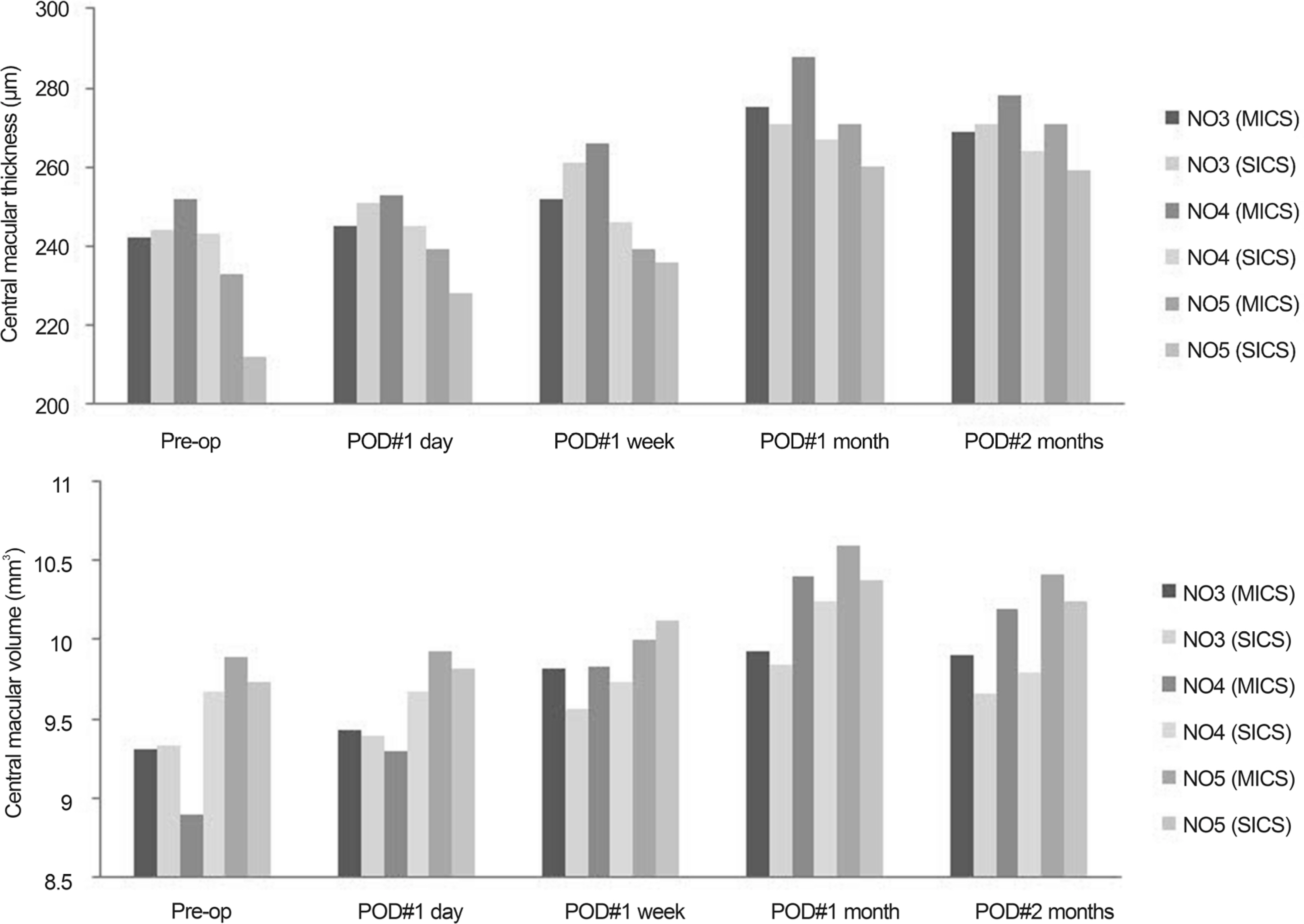
Figure 3. Subset analysis comparing central macular thickness and central macular volume between MICS and SICS group according to cataract grading (nuclear opacity of LOCS III) (student t-test p < 0.05*). POD = postoperative date; NO = nuclear opacity; SICS = small incision cataract surgery; MICS = microincision coaxial cataract surgery; LOCS III = lens opacities classification system III.
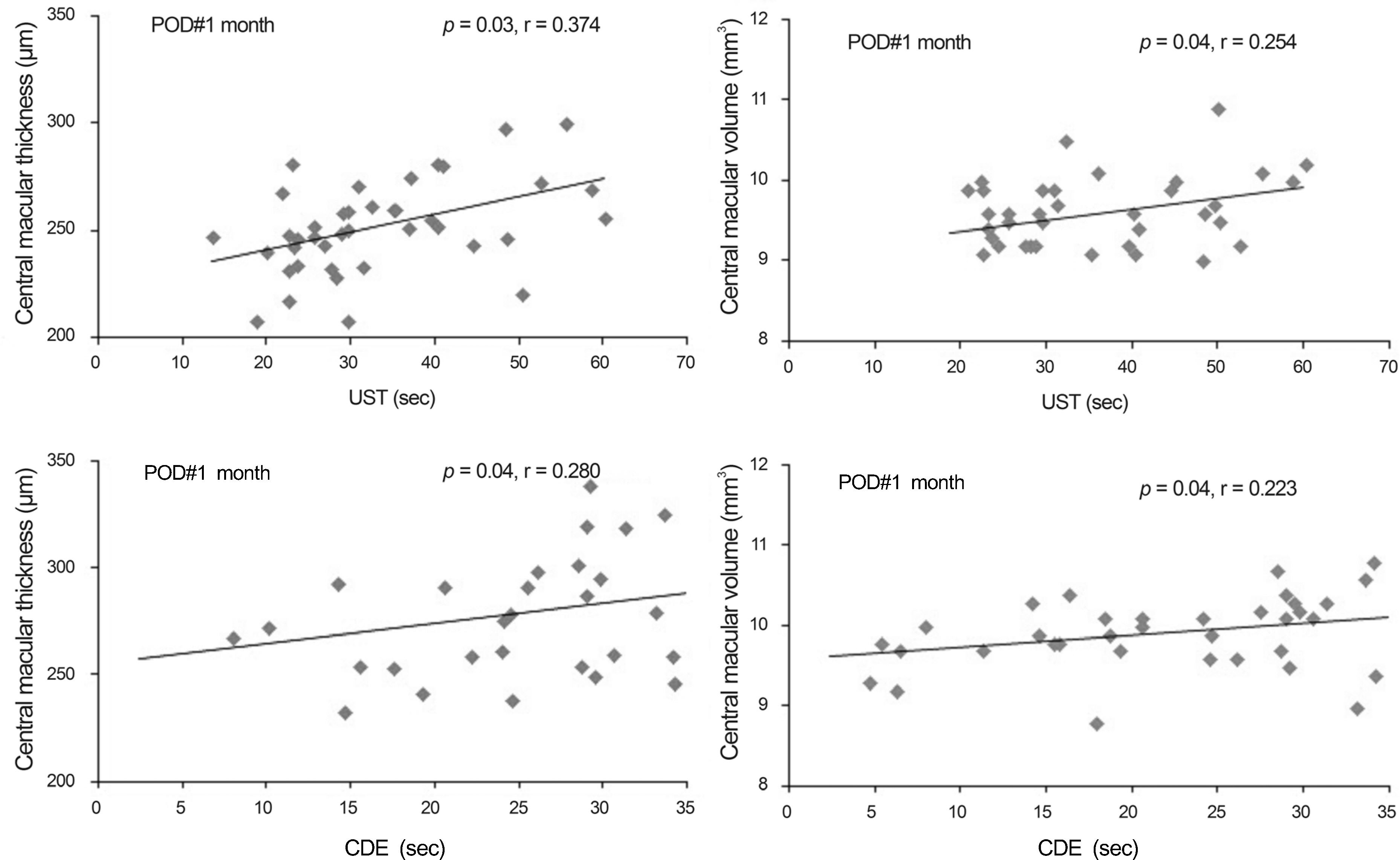
Figure 4. Correlation analysis of intraoperative factors (ultrasound time, cumulative dissipated energy) and central macular thickness, central macular volume, respectively (r = Pearson correlation coefficient). POD = postoperative date; UST = ultrasound time; CDE = cumulative dissipated energy.
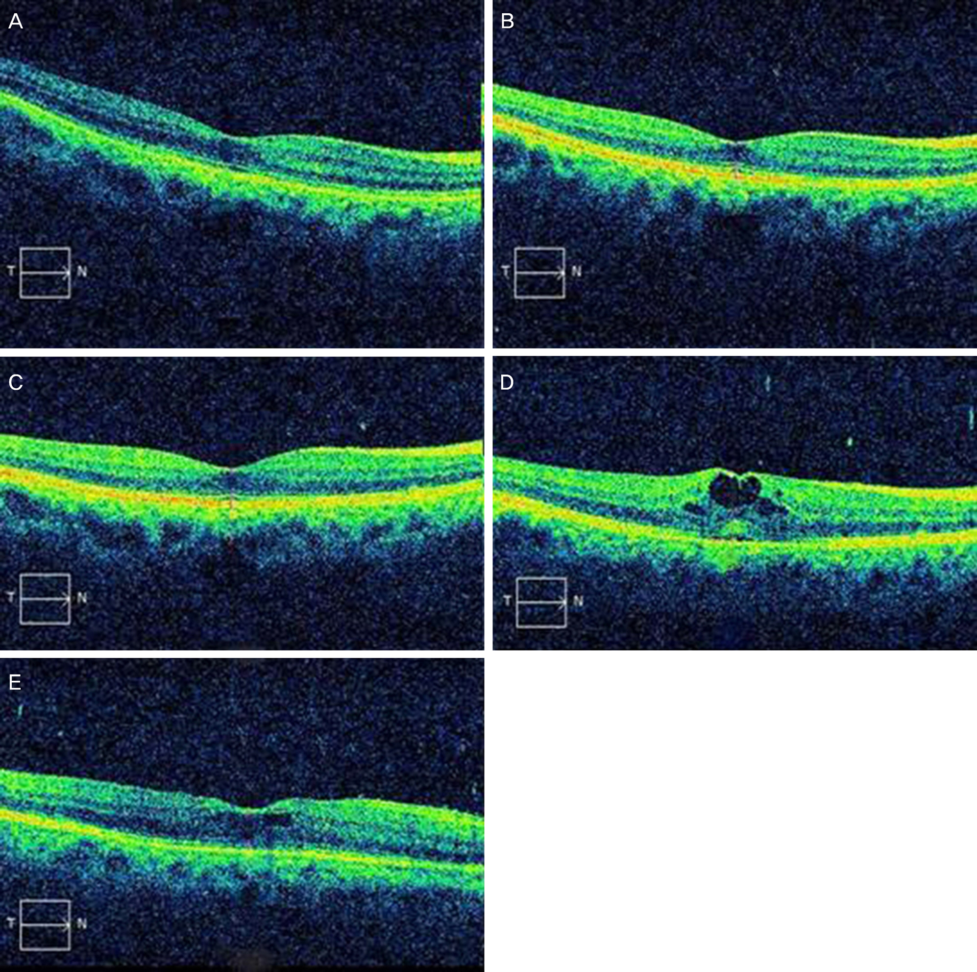
Figure 5. The optical coherence tomography (OCT) images of a patient with macular edema at preoperative (A) and postoperative 1 day (B), 1 week (C), 1 month (D), 2 months (E).
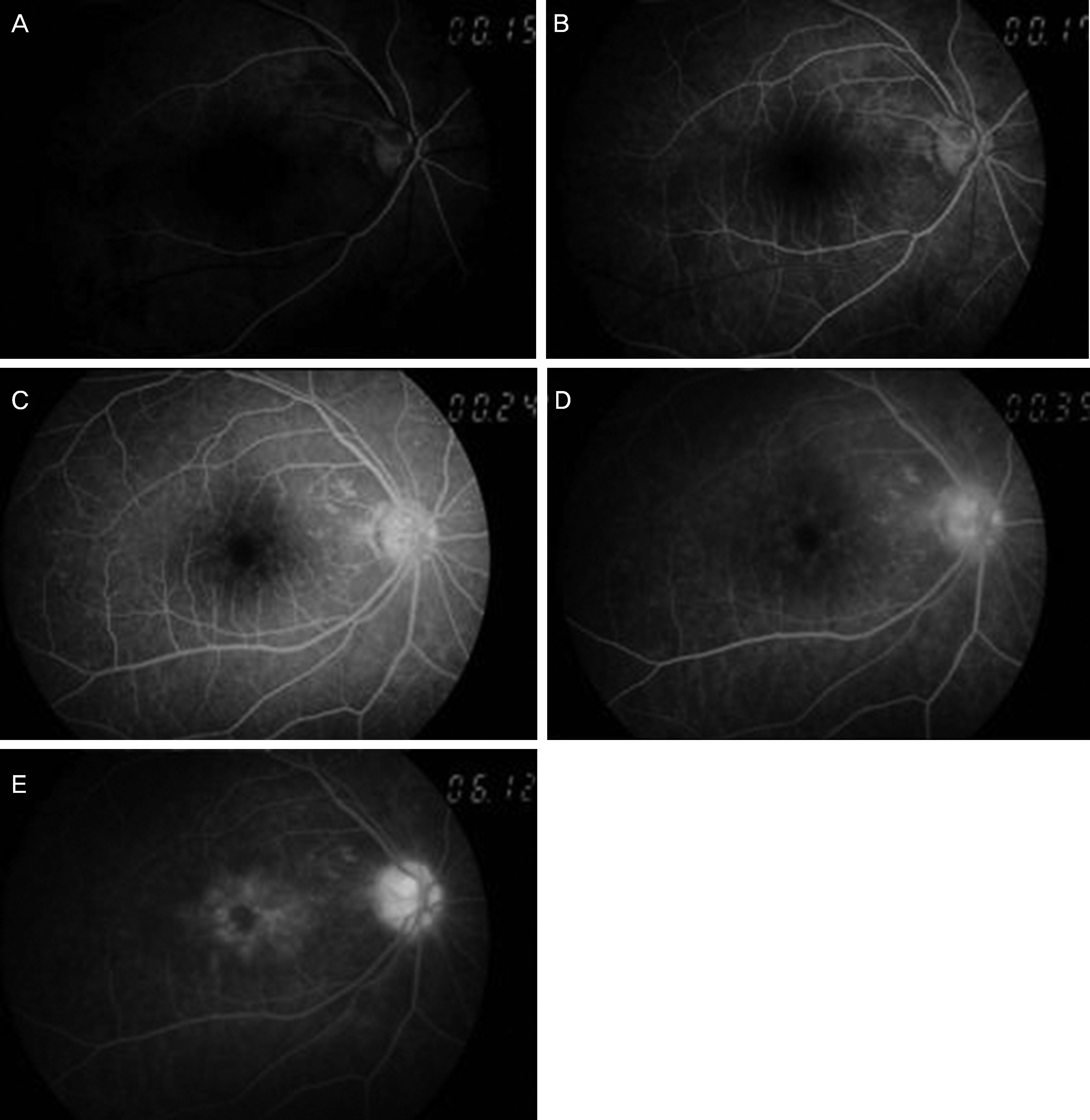
Figure 6. The FAG images of a patient with macular edema at postoperative 1 month: Arterial phase (A), A-V phase (B), venous phase (C), recirculation phase (D) and late phase (E). FAG = fluorecein angiography; A-V = arterial-venous.
Reference
-
References
1. Rossetti L, Autelitano A. Cystoid macular edema following cataract surgery. Curr Opin Ophthalmol. 2000; 11:65–72.
Article2. Rossetti L, Chaudhuri J, Dickersin K. Medical prophylaxis and treatment of cystoid macular edema after cataract surgery. The results of a meta-analysis. Ophthalmology. 1998; 105:397–405.3. Norregaard JC, Bernth-Petersen P, Bellan L, et al. Intraoperative clinical practice and risk of early complications after cataract extraction in the United States, Canada, Denmark, and Spain. Ophthalmology. 1999; 106:42–8.
Article4. Flesner P, Sander B, Henning V, et al. Cataract surgery on diabetic patients. A prospective evaluation of risk factors and complications. Acta Ophthalmol Scand. 2002; 80:19–24.
Article5. Hayashi K, Igarashi C, Hirata A, Hayashi H. Changes in diabetic macular oedema after phacoemulsification surgery. Eye (Lond). 2009; 23:389–96.
Article6. Kim SJ, Equi R, Bressler NM. Analysis of macular edema after cataract surgery in patients with diabetes using optical coherence tomography. Ophthalmology. 2007; 114:881–9.
Article7. Lobo CL, Faria PM, Soares MA, et al. Macular alterations after small-incision cataract surgery. J Cataract Refract Surg. 2004; 30:752–60.
Article8. Shepherd JR. Induced astigmatism in small incision cataract surgery. J Cataract Refract Surg. 1989; 15:85–8.
Article9. Dosso AA, Cottet L, Burgener ND, Di Nardo S. Outcomes of coaxial microincision cataract surgery versus conventional coaxial cataract surgery. J Cataract Refract Surg. 2008; 34:284–8.
Article10. Kim HJ, Kim JH, Lee DH. Endothelial cell damage in microincison cataract surgery and coaxial phacoemulsification. J Korean Ophthalmol Soc. 2007; 48:19–26.11. Can I, Takmaz T, Yildiz Y, et al. Coaxial, microcoaxial, and biaxial microincision cataract surgery: prospective comparative study. J Cataract Refract Surg. 2010; 36:740–6.12. Kurz S, Krummenauer F, Thieme H, Dick HB. Optical coherence tomography of macular thickness after biaxial vs coaxial microincision clear corneal cataract surgery. Eur J Ophthalmol. 2009; 19:990–7.
Article13. Findl O, Amon M, Petternel V, Kruger A. Early objective assessment of intraocular inflammation after phacoemulsification cataract surgery. J Cataract Refract Surg. 2003; 29:2143–7.
Article14. HOGAN MJ, KIMURA SJ, THYGESON P. Signs and symptoms of uveitis. I. Anterior uveitis. Am J Ophthalmol. 1959; 47:155–70.15. Kelman CD. Phaco-emulsification and aspiration. A new technique of cataract removal. A preliminary report. Am J Ophthalmol. 1967; 64:23–35.16. Dick HB. Controlled clinical trial comparing biaxial microincision with coaxial small incision for cataract surgery. Eur J Ophthalmol. 2012; 22:739–50.
Article17. Linebarger EJ, Hardten DR, Shah GK, Lindstrom RL. Phacoemulsification and modern cataract surgery. Surv Ophthalmol. 1999; 44:123–47.
Article18. Steinert RF, Brint SF, White SM, Fine IH. Astigmatism after small incision cataract surgery. A prospective, randomized, multicenter comparison of 4- and 6.5-mm incisions. Ophthalmology. 1991; 98:417–23. discussion 423-4.19. Kohnen T, Dick B, Jacobi KW. Comparison of the induced astigmatism after temporal clear corneal tunnel incisions of different sizes. J Cataract Refract Surg. 1995; 21:417–24.
Article20. Masket S, Wang L, Belani S. Induced astigmatism with 2.2- and 3.0-mm coaxial phacoemulsification incisions. J Refract Surg. 2009; 25:21–4.
Article21. Lobo C. Pseudophakic cystoid macular edema. Ophthalmologica. 2012; 227:61–7.
Article22. Ursell PG, Spalton DJ, Tilling K. Relation between postoperative blood-aqueous barrier damage and LOCS III cataract gradings following routine phacoemulsification surgery. Br J Ophthalmol. 1997; 81:544–7.
Article23. Ray S, D'Amico DJ. Pseudophakic cystoid macular edema. Semin Ophthalmol. 2002; 17:167–80.
Article24. Ruiz RS, Saatci OA. Visual outcome in pseudophakic eyes with clinical cystoid macular edema. Ophthalmic Surg. 1991; 22:190–3.
Article25. Kim JY, Song MH, Chung SK. Analysis of postoperative macular edema in cataract patients with diabetes using optical coherence tomography. J Korean Ophthalmol Soc. 2010; 51:340–6.
Article26. Mentes J, Erakgun T, Afrashi F, Kerci G. Incidence of cystoid macular edema after uncomplicated phacoemulsification. Ophthalmologica. 2003; 217:408–12.
Article27. Ursell PG, Spalton DJ, Whitcup SM, Nussenblatt RB. Cystoid macular edema after phacoemulsification: relationship to blood-aqueous barrier damage and visual acuity. J Cataract Refract Surg. 1999; 25:1492–7.
Article28. Ersoy L, Caramoy A, Ristau T, et al. Aqueous flare is increased in patients with clinically significant cystoid macular oedema after cataract surgery. Br J Ophthalmol. 2013; 97:862–5.
Article29. Kim EC, Byun YS, Kim MS. Microincision versus small-incision coaxial cataract surgery using different power modes for hard nuclear cataract. J Cataract Refract Surg. 2011; 37:1799–805.
Article30. Osher RH, Injev VP. Thermal study of bare tips with various system parameters and incision sizes. J Cataract Refract Surg. 2006; 32:867–72.
Article31. An TS, Park IW, Kwon SI. The changes in central macular thickness after cataract surgery in patients with diabetic retinopathy. J Korean Ophthalmol Soc. 2012; 53:1472–9.
Article32. Gharbiya M, Cruciani F, Cuozzo G, et al. Macular thickness changes evaluated with spectral domain optical coherence tomography after uncomplicated phacoemulsification. Eye (Lond). 2013; 27:605–11.
Article33. Kusbeci T, Eryigit L, Yavas G, Inan UU. Evaluation of cystoid macular edema using optical coherence tomography and fundus fluorescein angiography after uncomplicated phacoemulsification surgery. Curr Eye Res. 2012; 37:327–33.
Article34. Sahin M, Cingü AK, Gözüm N. Evaluation of cystoid macular edema using optical coherence tomography and fundus autofluorescence after uncomplicated phacoemulsification surgery. J Ophthalmol. 2013; 2013:376013.
- Full Text Links
-
- Actions
-
Cited
- CITED
-
- Close
- Share
-
- Similar articles
-
- Comparison of Surgically-induced Astigmatism after Combined Phacoemulsification and 23-Gauge Vitrectomy: 2.2-mm vs. 2.75-mm Cataract Surgery
- Comparison of Phacodynamic Effects on Postoperative Corneal Edema Between 2.8 mm and 2.2 mm Microcoaxial Torsional Phacoemulsification
- Comparison of Clinical Results between 2.2 mm and 2.8 mm Incision Cataract Surgery Using Ellips Ultrasound
- Endothelial Cell Damage in Microincison Cataract Surgery and Coaxial Phacoemulsification
- Induced Astigmatism and High-Order Aberrations after 1.8-mm, 2.2-mm and 3.0-mm Coaxial Phacoemulsification Incisions
