J Korean Ophthalmol Soc.
2014 Oct;55(10):1413-1417. 10.3341/jkos.2014.55.10.1413.
Calculated Brain CT Angiography Volumes of Lacrimal Glands in Normal Korean Orbits
- Affiliations
-
- 1Department of Ophthalmology, Dong-A University College of Medicine, Busan, Korea. hbahn@dau.ac.kr
- 2Department of Radiology, Dong-A University Hospital, Busan, Korea.
- KMID: 2216857
- DOI: http://doi.org/10.3341/jkos.2014.55.10.1413
Abstract
- PURPOSE
To determine the size range of lacrimal glands calculated from Brain CT angiography.
METHODS
A retrospective review of 107 CT scans of 214 orbits was performed. Aquaris Intuition Viewer software was used to calculate the volumes.
RESULTS
The mean volume of the lacrimal gland was 0.655 cm3 in right orbits and 0.595 cm3 in left orbits, 0.616 cm3 in men and 0.625 cm3 in women. There was a significant difference between right and left (p = 0.012) but no difference between men and women (p = 0.725). Linear regression analyses revealed that there was an inverse relationship between gland volume and age (Pearson r = -0.433, p < 0.001).
CONCLUSIONS
This is the first study to report the normal volume range of Korean lacrimal glands as measured by CT scans. A difference was detected in the volume between right and left lacrimal glands. The volume of the lacrimal gland decreased with age, and there were no gender differences.
MeSH Terms
Figure
-
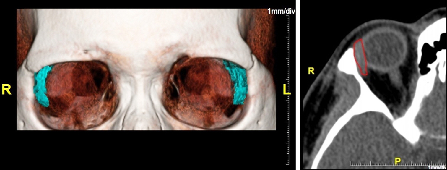
Figure 1. Coronal and axial view (brain CT angiography) on the Aquarlius intuition viewer with the entire lacrimal gland outlined. R = right; L = left; P = posterior.
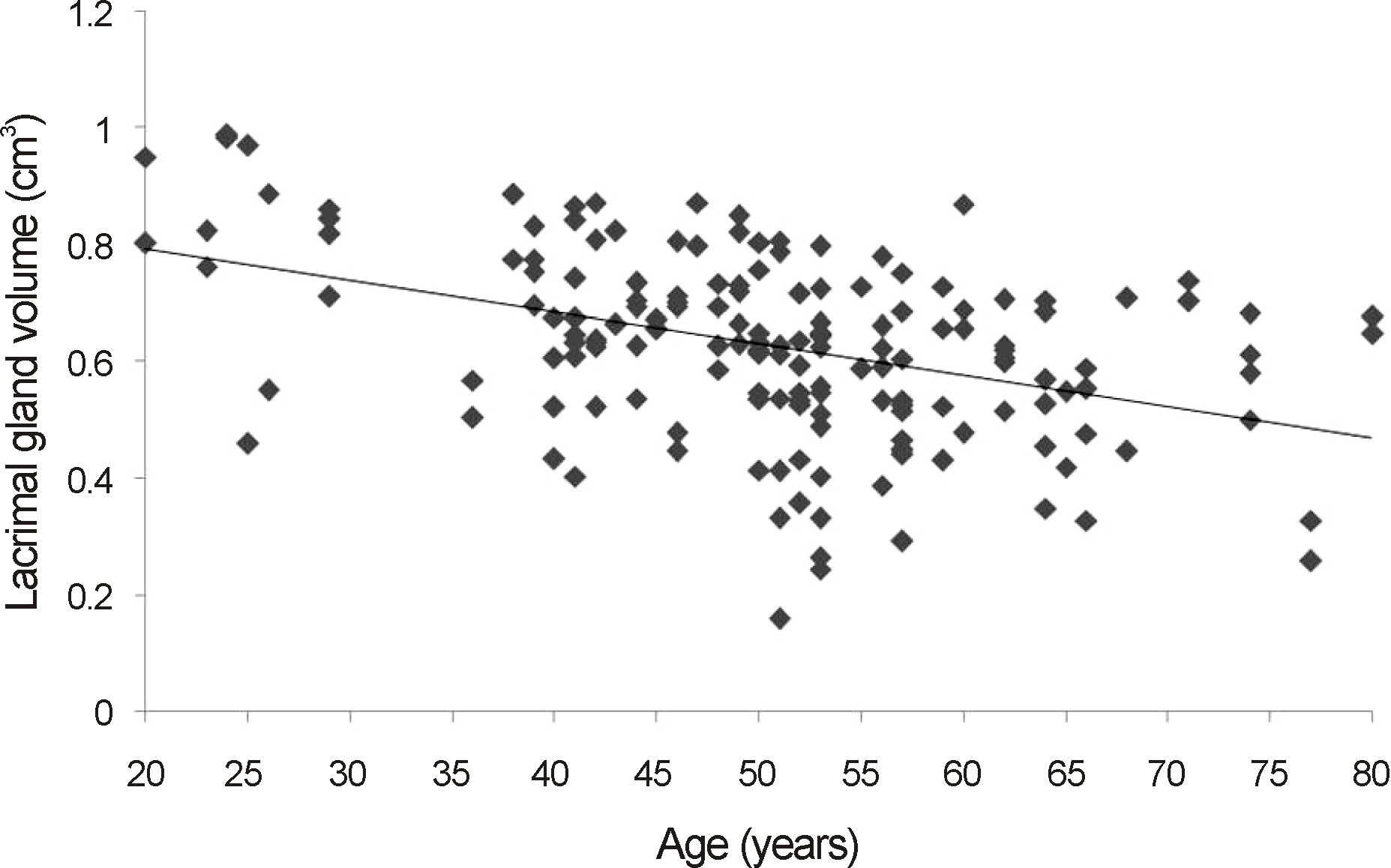
Figure 2. Lacrimal gland volume versus patients age.
Reference
-
References
1. Lorber M. Gross characteristics of normal human lacrimal glands. Ocul Surf. 2007; 5:13–22.
Article2. Bron AJ. Lacrimal streams: the demonstration of human lacrimal fluid secretion and the lacrimal ductules. Br J Ophthalmol. 1986; 70:241–5.
Article3. Hughes GK, Miszkiel KA. Imaging of the lacrimal gland. Semin Ultrasound CT MR. 2006; 27:476–91.
Article4. Jung WS, Ahn KJ, Park MR, et al. The radiological spectrum of orbital pathologies that involve the lacrimal gland and the lacrimal fossa. Korean J Radiol. 2007; 8:336–42.
Article5. Tamboli DA, Harris MA, Hogg JP, et al. Computed tomography dimensions of the lacrimal gland in normal Caucasian orbits. Ophthal Plast Reconstr Surg. 2011; 27:453–6.
Article6. Ueno H, Ariji E, Izumi M, et al. MR imaging of the lacrimal gland. Age-related and gender-dependent changes in size and structure. Acta Radiol. 1996; 37:714–9.7. Lee JS, Lee H, Kim JW, et al. Computed tomographic dimensions of the lacrimal gland in healthy orbits. J Craniofac Surg. 2013; 24:712–5.
Article8. Avetisov SE, Kharlap SI, Markosian AG, et al. [Ultrasound spatial clinical analysis of the orbital part of the lacrimal gland in health]. Vestn Oftalmol. 2006; 122:14–6.9. Lorber M. Gross characteristics of normal human lacrimal glands. Ocul Surf. 2007; 5:13–22.
Article10. Barrett JF, Keat N. Artifacts in CT recognition and avoidance. Radiographics. 2004; 24:1679–91.
Article11. Landis JR, Koch GG. The measurement of observer agreement for categorical data. Biometrics. 1977; 33:159–74.
Article12. Rootman J. Diseases of the orbit: a multidisciplinary approach. 2nd ed.Philadelphia: Lippincott Williams and Wilkins;2003. p. 344.13. Harris MA, Realini T, Hogg JP, Sivak-Callcott JA. CT dimensions of the lacrimal gland in Graves orbitopathy. Ophthal Plast Reconstr Surg. 2012; 28:69–72.
Article14. Forbes G, Gehring DG, Gorman CA, et al. Volume measurements of normal orbital structures by computed tomographic analysis. AJR Am J Roentgenol. 1985; 145:149–54.
Article15. Bingham CM, Castro A, Realini T, et al. Calculated CT volumes of lacrimal glands in normal Caucasian orbits. Ophthal Plast Reconstr Surg. 2013; 29:157–9.
Article16. Lorber M, Vidić B. Measurements of lacrimal glands from cadavers, with descriptions of typical glands and three gross variants. Orbit. 2009; 28:137–46.
Article17. Obata H. Anatomy and histopathology of the human lacrimal gland. Cornea. 2006; 25:S82–9.
Article18. Obata H, Yamamoto S, Horiuchi H, Machinami R. Histopathologic study of human lacrimal gland. Statistical analysis with special reference to aging. Ophthalmology. 1995; 102:678–86.
- Full Text Links
-
- Actions
-
Cited
- CITED
-
- Close
- Share
-
- Similar articles
-
- Calculated CT Volumes of Lacrimal Glands in Normal Korean Orbits
- Tympanometry and CT Measurement of Middle Ear Volumes in Patients with Unilateral Chronic Otitis Media
- Anatomical Relation between Anterior Ethmoidal Sinus and Lacrimal Sac Fossa on High Resolution CT
- A Case of Mikulicz's Disease
- Anatomical Study of Lacrimal Passage using Computed Tomography
