Changes in Higher-Order Aberrations after Penetrating Keratoplasty
- Affiliations
-
- 1Department of Ophthalmology, Chonnam National University Hospital, Chonnam National University Medical School, Gwangju, Korea. kcyoon@chonnam.ac.kr
- KMID: 2215950
- DOI: http://doi.org/10.3341/jkos.2012.53.8.1088
Abstract
- PURPOSE
To evaluate the changes in corneal higher-order aberrations (HOAs) after penetrating keratoplasty (PKP) and to investigate the factors affecting the changes in corneal HOAs.
METHODS
Forty-eight eyes of 48 patients with three different underlying diseases who underwent PKP were retrospectively reviewed. The changes in corneal higher-order aberrations (total root mean square (RMS), coma, trefoil, and spherical aberration) were evaluated with Pentacam (Oculus Inc., Dutenhofen, Germany) at 1, 3, 6, 12, and 18 months postoperatively. Sex, age, underlying disease, suturing method, trephine size of donor, and suture state were analyzed as factors affecting the HOAs changes.
RESULTS
Total RMS values in the PKP eyes were 1.67 +/- 0.49 microm at 1 month, 1.63 +/- 0.49 microm at 3 months, 1.64 +/- 0.51 microm at 6 months, 1.61 +/- 0.50 microm at 12 months, and 1.79 +/- 0.40 microm at 18 months after surgery (p < 0.01). The values were higher compared with those in the control eyes. Sex, donor trephine size and suture method did not correlate with the HOAs changes. The HOAs in the keratoconus group were higher than those in the corneal opacity group or bullous keratopathy group. At 18 months, HOAs in the suture removed group were higher than those in the suture-remaining group.
CONCLUSIONS
Corneal HOAs after PKP were persistently higher than those in the control eyes. The HOAs were higher in patients with keratoconus and in the suture-removed group.
MeSH Terms
Figure
-
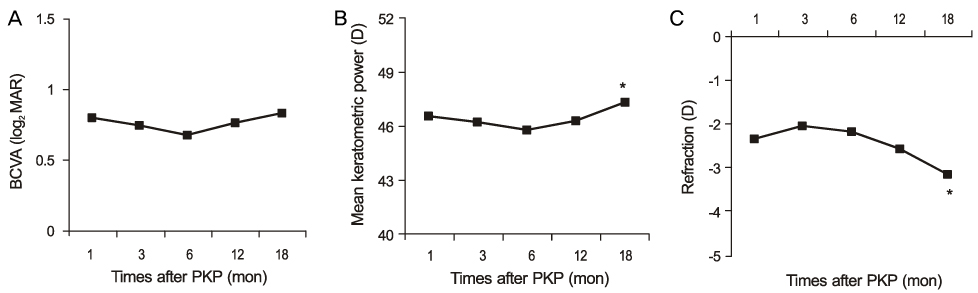
Figure 1 Changes in best corrected visual acuity (A), mean keratometric power (B), spherical equivalent (C) after penetrating keratoplasty. D = diopter. *p < 0.05 compared with other periods, repeated measures ANOVA.
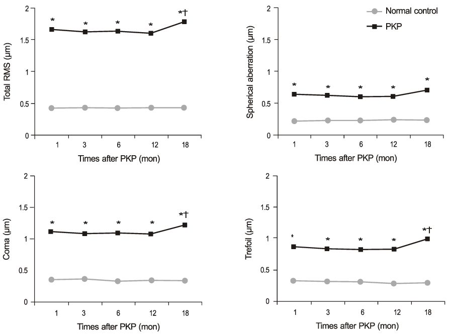
Figure 2 The changes of higher-order aberrations in the penetrating keratoplasty and control group. RMS = root mean square; PKP = penetrating keratoplasty. *p < 0.01 compared with the control group, paired t-test; †p < 0.05 compared with other periods, repeated measures ANOVA.

Figure 3 Correlation between age and higher-order aberrations including total RMS, coma, trefoil and spherical aberration at 12 months after penetrating keratoplasty. RMS = root mean square.

Figure 4 Comparison of the higher-order aberrations after penetrating keratoplasty according to the original diseases. RMS = root mean square. *p < 0.05 compared with the keratitis group; †p < 0.05 compared with the bullous keratopathy group, Kruskall-W allis test followed by Mann-Whitney U test with Bonfferoni correction.
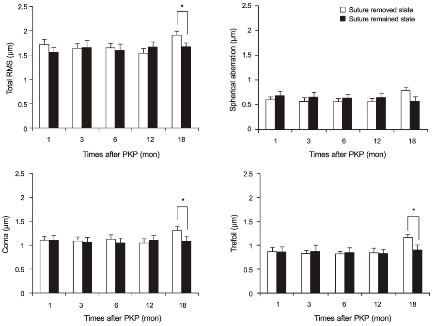
Figure 5 Comparison of the higher-order aberrations after penetrating keratoplasty according to the suture state at postoperative 18 months. RMS = root mean square. *p < 0.05 campared with the group that suture remained, Mann-Whitney U test.
Reference
-
1. Ing JJ, Ing HH, Nelson LR, et al. Ten-year postoperative results of penetrating keratoplasty. Ophthalmology. 1998. 105:1855–1865.2. Thompson RW Jr, Price MO, Bowers PJ, Price FW Jr. Long-term graft survival after penetrating keratoplasty. Ophthalmology. 2003. 110:1396–1402.3. Jung KI, Choi JA, Na KS, et al. Comparison of outcomes of femtosecond laser-assisted keratoplasty and conventional penetrating keratoplasty. J Korean Ophthalmol Soc. 2010. 51:1054–1063.4. Akcay L, Kaplan AT, Kandemir B, et al. Toric intraocular collamer lens for high myopic astigmatism after penetrating keratoplasty. J Cataract Refract Surg. 2009. 35:2161–2163.5. Donnenfeld ED, Kornstein HS, Amin A, et al. Laser in situ keratomileusis for correction of myopia and astigmatism after penetrating keratoplasty. Ophthalmology. 1999. 106:1966–1974.6. Pantanelli S, MacRae S, Jeong TM, Yoon G. Characterizing the wave aberration in eyes with keratoconus or penetrating keratoplasty using a high-dynamic range wavefront sensor. Ophthalmology. 2007. 114:2013–2021.7. Pesudovs K, Coster DJ. Penetrating keratoplasty for keratoconus: the nexus between corneal wavefront aberrations and visual performance. J Refract Surg. 2006. 22:926–931.8. Yagci A, Egrilmez S, Kaskaloglu M, Egrilmez ED. Quality of vision following clinically successful penetrating keratoplasty. J Cataract Refract Surg. 2004. 30:1287–1294.9. Shah S, Naroo S, Hosking S, et al. Nidek OPD-scan analysis of normal, keratoconic, and penetrating keratoplasty eyes. J Refract Surg. 2003. 19:2 Suppl. S255–S259.10. McLaren JW, Patel SV, Bourne WM, Baratz KH. Corneal wavefront errors 24 months after deep lamellar endothelial keratoplasty and penetrating keratoplasty. Am J Ophthalmol. 2009. 147:959–965.11. Miranda MA, O'Donnell C, Radhakrishnan H. Repeatability of corneal and ocular aberration measurements and changes in aberrations over one week. Clin Exp Optom. 2009. 92:253–266.12. Muftuoglu O, Prasher P, Bowman RW, et al. Corneal higher-order aberrations after Descemet's stripping automated endothelial keratoplasty. Ophthalmology. 2010. 117:878–884.13. Choi SH, Lee YW, Kim HM, et al. Epidemiologic studies of keratoplasty in Korea. J Korean Ophthalmol Soc. 2006. 47:538–547.14. Farid M, Kim M, Steinert RF. Results of penetrating keratoplasty performed with a femtosecond laser zigzag incision initial report. Ophthalmology. 2007. 114:2208–2212.15. Ignacio TS, Nguyen TB, Chuck RS, et al. Top hat wound configuration for penetrating keratoplasty using the femtosecond laser: a laboratory model. Cornea. 2006. 25:336–340.16. Webber SK, Lawless MA, Sutton GL, Rogers CM. LASIK for post penetrating keratoplasty astigmatism and myopia. Br J Ophthalmol. 1999. 83:1013–1018.17. Tahzib NG, Cheng YY, Nuijts RM. Three-year follow-up analysis of Artisan toric lens implantation for correction of postkeratoplasty ametropia in phakic and pseudophakic eyes. Ophthalmology. 2006. 113:976–984.18. Arriola-Villalobos P, Díaz-Valle D, Güell JL, et al. Intrastromal corneal ring segment implantation for high astigmatism after penetrating keratoplasty. J Cataract Refract Surg. 2009. 35:1878–1884.19. Hjortdal JØ, Ehlers N. Treatment of post-keratoplasty astigmatism by topography supported customized laser ablation. Acta Ophthalmol Scand. 2001. 79:376–380.20. Hindman HB, McCally RL, Myrowitz E, et al. Evaluation of deep lamellar endothelial keratoplasty surgery using scatterometry and wavefront analyses. Ophthalmology. 2007. 114:2006–2012.21. Son JH, Tchah H, Kim YJ. Suture tension adjustment of single running suture in penetrating keratoplasty. J Korean Ophthalmol Soc. 1993. 34:198–201.22. Goren MB, Dana MR, Rapuano CJ, et al. Corneal topography after selective suture removal for astigmatism following keratoplasty. Ophthalmic Surg Lasers. 1997. 28:208–214.23. Solano JM, Hodge DO, Bourne WM. Keratometric astigmatism after suture removal in penetrating keratoplasty: double running versus single running suture techniques. Cornea. 2003. 22:716–720.24. Kim KE, Joo CK. Changes in astigmatism after suture removal in penetrating keratoplasty. J Korean Ophthalmol Soc. 2003. 44:284–288.25. Lin DT, Wilson SE, Reidy JJ, et al. Topographic changes that occur with 10-0 running suture removal following penetrating keratoplasty. Refract Corneal Surg. 1990. 6:21–25.26. Shimazaki J, Tsubota K. Analysis of videokeratography after penetrating keratoplasty: topographic characteristics and effects of removing running sutures. Ophthalmology. 1997. 104:2077–2084.27. Amano S, Amano Y, Yamagami S, et al. Age-related changes in corneal and ocular higher-order wavefront aberrations. Am J Ophthalmol. 2004. 137:988–992.28. Sicam VA, Dubbelman M, van der Heijde RG. Spherical aberration of the anterior and posterior surfaces of the human cornea. J Opt Soc Am A Opt Image Sci Vis. 2006. 23:544–549.29. Guirao A, Redondo M, Artal P. Optical aberrations of the human cornea as a function of age. J Opt Soc Am A Opt Image Sci Vis. 2000. 17:1697–1702.30. Wang L, Dai E, Koch DD, Nathoo A. Optical aberrations of the human anterior cornea. J Cataract Refract Surg. 2003. 29:1514–1521.31. Spadea L, Cifariello F, Bianco G, Balestrazzi E. Long-term results of penetrating keratoplasty using a single or double running suture technique. Graefes Arch Clin Exp Ophthalmol. 2002. 240:415–419.32. Maeda N, Fujikado T, Kuroda T, et al. Wavefront aberrations measured with Hartmann-Shack sensor in patients with keratoconus. Ophthalmology. 2002. 109:1996–2003.33. Atchison DA, Mathur A, Read SA, et al. Peripheral ocular aberrations in mild and moderate keratoconus. Invest Ophthalmol Vis Sci. 2010. 51:6850–6857.34. de la Paz MF, Sibila GR, Montenegro G, et al. Wedge resection for high astigmatism after penetrating keratoplasty for keratoconus: refractive and histopathologic changes. Cornea. 2010. 29:595–600.35. Patel SV, Malta JB, Banitt MR, et al. Recurrent ectasia in corneal grafts and outcomes of repeat keratoplasty for keratoconus. Br J Ophthalmol. 2009. 93:191–197.36. Seitz B, Langenbucher A, Küchle M, Naumann GO. Impact of graft diameter on corneal power and the regularity of postkeratoplasty astigmatism before and after suture removal. Ophthalmology. 2003. 110:2162–2167.37. Filatov V, Steinert RF, Talamo JH. Postkeratoplasty astigmatism with single running suture or interrupted sutures. Am J Ophthalmol. 1993. 115:715–721.38. Karabatsas CH, Cook SD, Figueiredo FC, et al. Combined interrupted and continuous versus single continuous adjustable suturing in penetrating keratoplasty: a prospective, randomized study of induced astigmatism during the first postoperative year. Ophthalmology. 1998. 105:1991–1998.39. Raecker ME, Erie JC, Patel SV, et al. Long-term keratometric changes after penetrating keratoplasty for keratoconus and Fuchs Endothelial dystrophy. Am J Ophthalmol. 2009. 147:227–233.
- Full Text Links
-
- Actions
-
Cited
- CITED
-
- Close
- Share
-
- Similar articles
-
- Cataract Extraction after Penetrating Keratoplasty
- Astigmatism after Penetrating Keratoplasty According to Suture Methods
- Clinical Evaluation of the Glaucoma after Penetrating Keratoplasty
- Patho-Physiology of Penetrating Keratoplasty
- A Case of Resolved Massive Suprachoroidal Hemorrhage after Sclerotomy in Penetrating Keratoplasty Patient
