Clinical Results of Intacs(R) Ring Implantation in Keratoconus or Keratectasia
- Affiliations
-
- 1Department of Ophthalmology, Seoul National University College of Medicine, Seoul, Korea. Kmk9@snu.ac.kr
- 2Laboratory of Ocular Regenerative Medicine and Immunology, Seoul Artificial Eye Center, Seoul National University Hospital Clinical Research Institute, Seoul, Korea.
- KMID: 2215699
- DOI: http://doi.org/10.3341/jkos.2015.56.4.499
Abstract
- PURPOSE
To report the clinical results after the implantation of intrastromal corneal ring segments (Intacs(R)) for the correction of keratoconus or keratectasia.
METHODS
This retrospective study was comprised of 16 eyes treated by insertion of intrastromal corneal ring and 30 eyes treated by penetrating keratoplasty (PKP) who were diagnosed with keratoconus or keratectasia. Visual acuity, refractive outcome, keratometric values were evaluated before and at 3 months, 6 months, and 12 months postoperatively. In addition, the implanted ring segment depth was measured by anterior segment optical coherence tomography and the results were compared based on the depth of the ring.
RESULTS
Twelve months after treatment, best corrected visual acuity (BCVA) was log MAR 0.32 at the ring group and log MAR 0.20 at the PKP group. BCVA change was larger at the PKP group than the ring group. Postoperative keratometric value was smaller at the ring group than at the PKP group. 3 mm irregular astigmatism was larger at the ring group than at the PKP group. The shallowly implanted ring group had a larger effect than the deeply implanted ring group.
CONCLUSIONS
Intrastromal corneal ring segment implantation appears to be effective in improving the visual acuity and refractive outcome, although it cannot substitute for PKP.
MeSH Terms
Figure
-

Figure 1. Corneal topography (A) and pachymetric map of anterior segment OCT (B) were used to design axis and stromal depth of the ring location. OCT = optical coherence tomography; S = superior; I = inferior; T = temporal; N = nasal.

Figure 2. Anterior to the ring (A) and posterior to the ring (B) portion of corneal thickness was gathered using anterior segment optical coherence tomography. Deep group is defined if (A) is same or longer than doubled (B). Shallow group is defined if (A) is shorter than doubled (B).
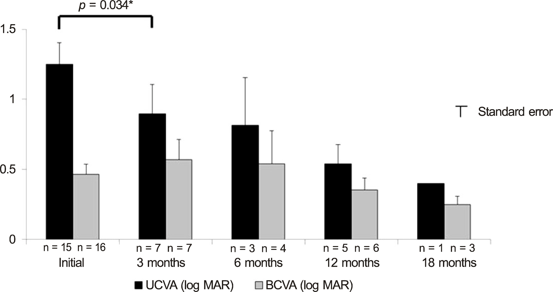
Figure 3. Changes in UCVA and BCVA after Intacs® implantation. The BCVA improved at postoperative 3 months. UCVA = uncorrected visual acuity; BCVA = best corrected visual acuity. * Wilcoxon signed rank test.
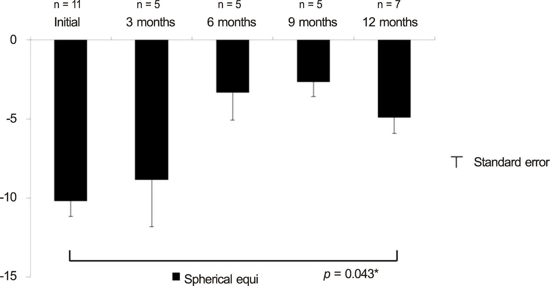
Figure 4. Changes in spherical equivalent (SE) after intacs implantation. The SE was improved at postoperative 12 months. * Wilcoxon signed rank test.

Figure 5. Manifest astigmatism before surgery reduced on average following surgery. Each data point represents the astigmatism component of a power vector for one eye, referenced to the spectacle plane. J0 = a Jackson crossed cylinder of power axes at 90 degrees and 180 degrees; J45 = a Jackson crossed cylinder of power axes at 45 degrees and 135 degrees. Power vector analysis (S = spherical diopters; C = cylindrical diopters; α = axis (degree); J0 = (-C/2) cos(2α); J45 = (-C/2) sin(2α).
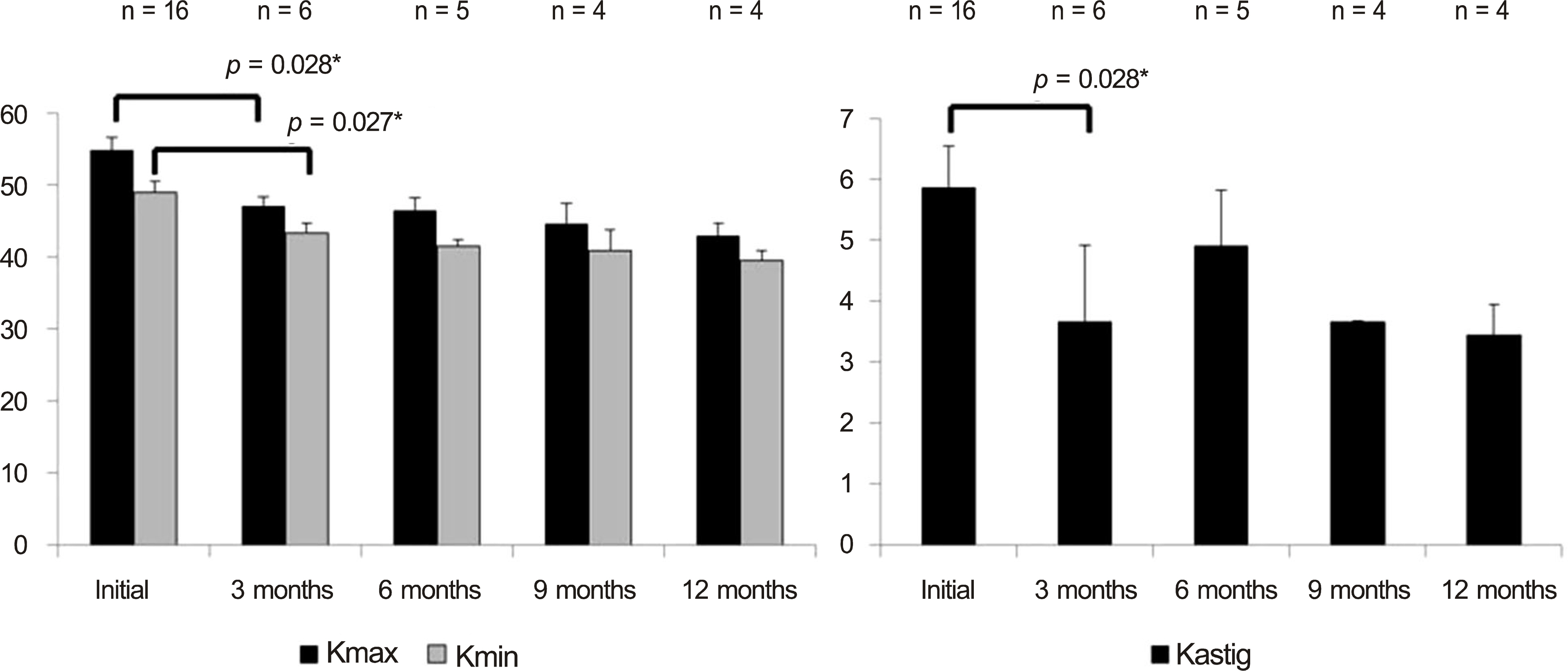
Figure 6. Changes in Kmax, Kmin and Kastig after intacs implantation. All the values improved at postoperative 3 months. Kmax = Kmaximum; Kmin = Kminimun; Kastig = Kastigmatism. * Wilcoxon signed rank test.
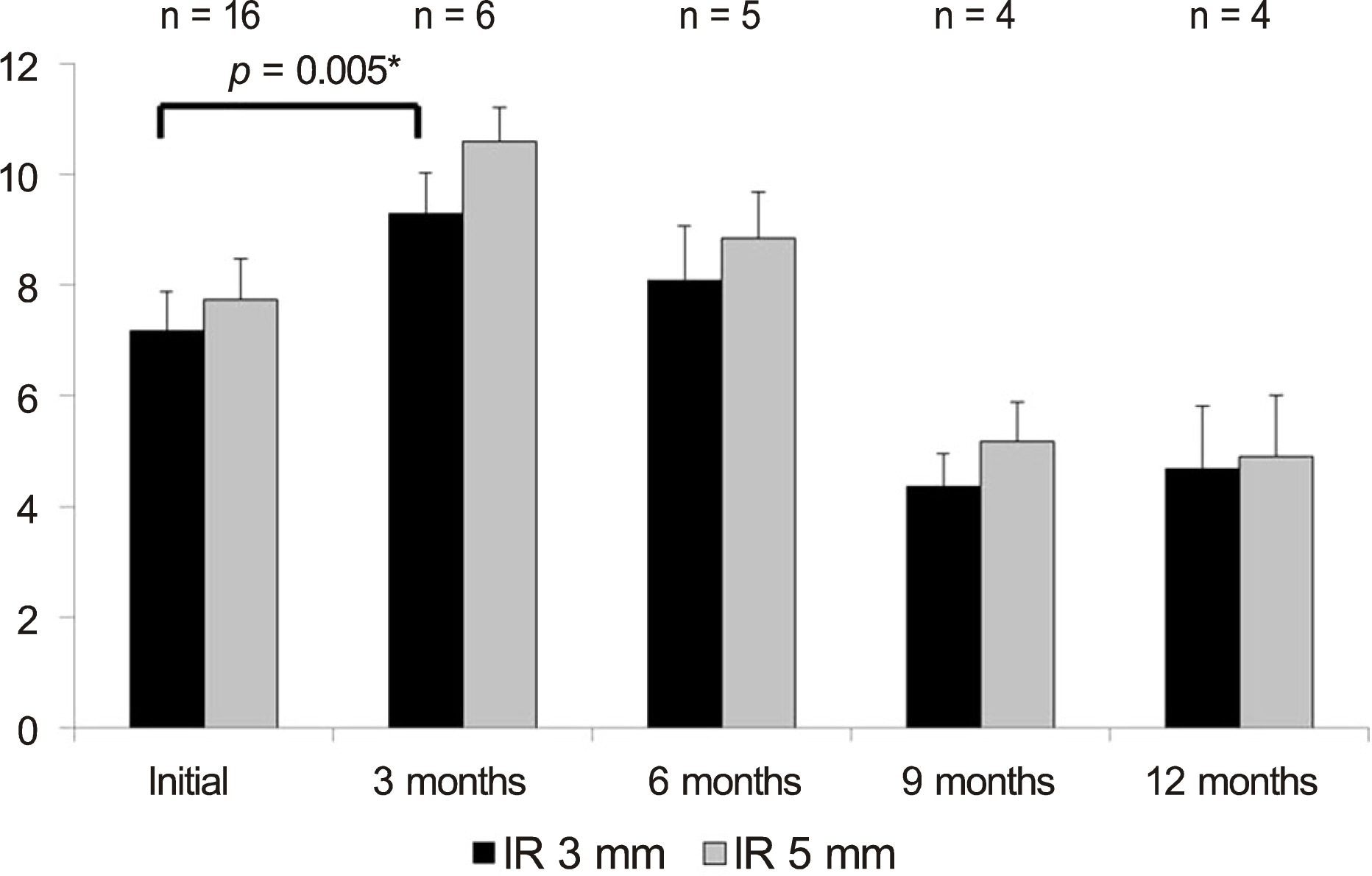
Figure 7. Changes in the irregular astigmatism of 3-mm and 5-mm area improved at postoperative 3 months. IR = irregular astigmatism. * Wilcoxon signed rank test.
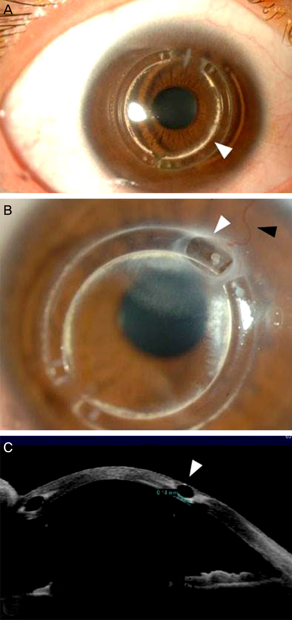
Figure 8. Complications of intrastromal corneal ring insertion. Marginal precipitation of the ring (white arrow head) is observed (A). Overlying stromal thinning (white arrow head) developed after new vessel formation (black arrow head) (B and C).
Reference
-
References
1. Kenney MC, Brown DJ, Rajeev B. Everett Kinsey lecture. The elu-sive causes of keratoconus: a working hypothesis. CLAO J. 2000; 26:10–3.2. Troutman RC, Gaster RN. Surgical advances and results of keratoconus. Am J Ophthalmol. 1980; 90:131–6.
Article3. Wagoner MD, Ba-Abbad R, Al-Mohaimeed M, et al. Postoperative complications after primary adult optical penetrating keratoplasty: prevalence and impact on graft survival. Cornea. 2009; 28:385–94.
Article4. Wollensak G, Spoerl E, Reber F, Seiler T. Keratocyte cytotoxicity of riboflavin/UVA-treatment in vitro. Eye (Lond). 2004; 18:718–22.
Article5. Godoy CS, Wahab SA, Moreira H, et al. [Analysis of corneal curvature alteration following intrastromal corneal ring implantation: experimental study in rabbits]. Arq Bras Oftalmol. 2007; 70:303–11.6. Kim EJ, Koo SH, Lee GJ, et al. The clinical results of Intacs(R) ring implantation by manual tunnel creation in patients with keratoconus. J Korean Ophthalmol Soc. 2012; 53:1756–65.7. Kim HS, Lee TH, Lee KH. Intracorneal ring segment implantation for the management of keratoconus: short-term safety and efficacy. J Korean Ophthalmol Soc. 2009; 50:1505–9.
Article8. Torquetti L, Ferrara G, Almeida F, et al. Intrastromal corneal ring segments implantation in patients with keratoconus: 10-year fol-low-up. J Refract Surg. 2014; 30:22–6.
Article9. Siganos D, Ferrara P, Chatzinikolas K, et al. Ferrara intrastromal corneal rings for the correction of keratoconus. J Cataract Refract Surg. 2002; 28:1947–51.
Article10. McMahon TT, Szczotka-Flynn L, Barr JT, et al. A new method for grading the severity of keratoconus: the Keratoconus Severity Score (KSS). Cornea. 2006; 25:794–800.11. Thibos LN, Horner D. Power vector analysis of the optical outcome of refractive surgery. J Cataract Refract Surg. 2001; 27:80–5.
Article12. Fleming JF, Wan WL, Schanzlin DJ. The theory of corneal curvature change with the Intrastromal Corneal Ring. CLAO J. 1989; 15:146–50.13. Twa MD, Karpecki PM, King BJ, et al. One-year results from the phase III investigation of the KeraVision Intacs. J Am Optom Assoc. 1999; 70:515–24.14. Colin J, Cochener B, Savary G, Malet F. Correcting keratoconus with intracorneal rings. J Cataract Refract Surg. 2000; 26:1117–22.
Article15. Torquetti L, Berbel RF, Ferrara P. Long-term follow-up of intrastromal corneal ring segments in keratoconus. J Cataract Refract Surg. 2009; 35:1768–73.
Article16. Coskunseven E, Kymionis GD, Tsiklis NS, et al. One-year results of intrastromal corneal ring segment implantation (KeraRing) using femtosecond laser in patients with keratoconus. Am J Ophthalmol. 2008; 145:775–9.
Article17. Piñero DP, Alio JL, El Kady B, et al. Refractive and aberrometric outcomes of intracorneal ring segments for keratoconus: mechan-ical versus femtosecond-assisted procedures. Ophthalmology. 2009; 116:1675–87.
Article18. Coskunseven E, Jankov MR 2nd, Hafezi F, et al. Effect of treatment sequence in combined intrastromal corneal rings and corneal collagen crosslinking for keratoconus. J Cataract Refract Surg. 2009; 35:2084–91.
Article19. MacIntyre R, Chow SP, Chan E, Poon A. Long-term outcomes of deep anterior lamellar keratoplasty versus penetrating keratoplasty in Australian keratoconus patients. Cornea. 2014; 33:6–9.
Article
- Full Text Links
-
- Actions
-
Cited
- CITED
-
- Close
- Share
-
- Similar articles
-
- Intracorneal Ring Segment Implantation for the Management of Keratoconus: Short-Term Safety and Efficacy
- The Clinical Results of Intacs(R) Ring Implantation by Manual Tunnel Creation in Patients with Keratoconus
- The Clinical Results of Intrastromal Corneal Ring Segment Implantation Using a Femtosecond Laser in Keratectasia
- A Case of Fungal Keratitis after Intracorneal Ring Segment Implantation for Keratoconus
- A Case of Keratectasia 5 Years after Photorefractive Keratectomy
